
13.1 POJA-L4552+4553
Myocardial fibrosis (after infarction, human)
13.1 POJA-L4552+4553
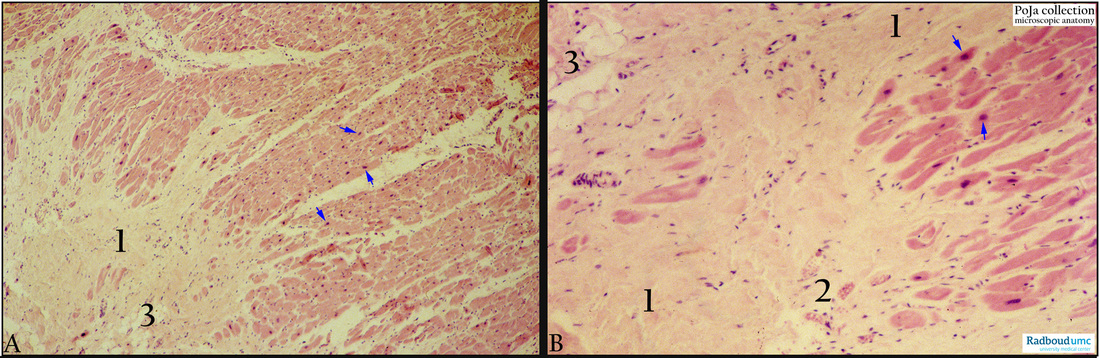
Title: Myocardial fibrosis (after infarction, human)
Description:
(A, B): Hypertrophic heart, myocardial fibrosis, Haematoxylin-eosin. After a myocardial infarction within 5-10 days macrophages remove necrotic cardiomyocytes and the damaged area is progressively replaced by the ingrowth of vascularised granulation tissue (2-4 weeks). The vital myocytes close to the infarction area show vacuolar dystrophy due to damage by hypoxia. Vacuolisation of myocytes can result in myolysis and will be repaired by reactive connective tissue proliferation. The fibrosis progresses and the myocytes are gradually replaced by hyalinised connective tissue (1). In most instances scarring is well advanced by the end of the 6th week. Within the scar small groups of dystrophic vacuolised myocardial cells (2) remain. Additional findings are the presence of accumulated adipocytes (so-called lipomatosis) (3) which can be detected in obese and in elderly people. Hypertrophic myocytes with conspicuous nuclei are also detectable (arrows).
Background: Summary of sequelae during infarction. Myocardial infarctions show ischaemic coagulation necrosis due to lack of oxygen i.e. obstruction of the coronary arteries (e.g. arteriosclerosis, thrombosis). After 1-3 days coagulation necrosis occurs with loss of nuclei including striations and interstitial infiltrate of neutrophils. Prominent at 2-3 days necrotic muscle cells with acute inflammation are present. In 3-7 days start of beginning disintegration of necrotic myofibres, dying neutrophils and early phagocytosis of dead cells by macrophages at the infarct boundary. After 7-10 days macrophages removed the necrotic myocytes and the damaged zone is progressively replaced by the ingrowth of fibrovascular granulation tissue at the margins of the infarction. In 10-14 days well-established granulation tissue with new blood vessels and collagen deposition are observed and after 2-8 weeks increased collagen deposition with decreased vascularity and cellularity. At the end of the 6th week scarring is well advanced and after more than 2 months a dense collagenous scar remains. Once a laesion is completely healed it is hardly possible to distinguish between an eight-week-old laesion and a ten-year-old one. (Partly adapted from ref. Robbins & Cotran Pathological Base of Disease, 8th ed. 2014, W.B. Saunders Company, ISBN 978-1-4160-3121-5). (http://www.us.elsevierhealth.com/robbins-pathology/robbins-cotran-pathologic-basis-of-disease-hardcover/9781416031215/
Keywords/Mesh: cardiovascular system, heart, ventricle, fibrosis, necrosis, lipomatosis, infarction, myocardial infarction, myolysis, histology, POJA collection