
12.1.6 POJA-L3579+3584+
3631.
Lacrimal gland 2
12.1.6 POJA-L3579+3584+3631
Title: Lacrimal gland 2
Description:
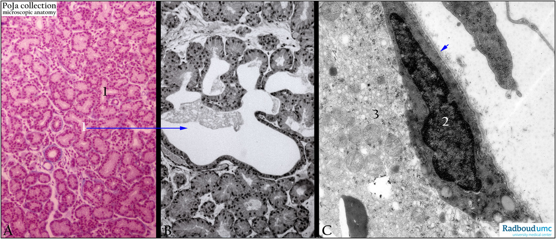
Lacrimal gland: (A): stain Azan, human. (B): Lacrimal gland, stain hematoxylin-eosin (black and white print), rat. (C): Myoepithelial cell,
electron micrograph, rat. The lacrimal gland is a branched tubuloacinar serous gland (A) with intralobular ducts (1).
(B) The acini often widen into alveolar-like structures.
The acinar structures are surrounded by myoepithelial cells (C, 2), these cells are located just below the basal lamina (arrow).
(C, 3) Part of a gland cell with organelles and one secretion droplet.
Keywords/Mesh: eye, lacrimal gland, myoepithelial cell, histology, electron microscopy, POJA collection
Title: Lacrimal gland 2
Description:
Lacrimal gland: (A): stain Azan, human. (B): Lacrimal gland, stain hematoxylin-eosin (black and white print), rat. (C): Myoepithelial cell,
electron micrograph, rat. The lacrimal gland is a branched tubuloacinar serous gland (A) with intralobular ducts (1).
(B) The acini often widen into alveolar-like structures.
The acinar structures are surrounded by myoepithelial cells (C, 2), these cells are located just below the basal lamina (arrow).
(C, 3) Part of a gland cell with organelles and one secretion droplet.
Keywords/Mesh: eye, lacrimal gland, myoepithelial cell, histology, electron microscopy, POJA collection