
4.2.1 POJA-L3748+3749. Portal triad in the liver (rat)
4.2.1 POJA-L3748+3749
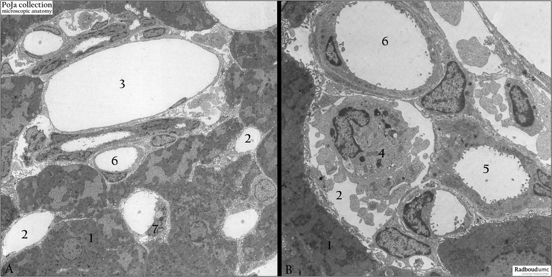
Title: Portal triad in the liver (rat)
Description: (A, B) Electron micrographs of perfused liver showing components of the portal triad in the liver.
(1) Liver parenchymal cells.
(2) Sinusoid space.
(3) Branch of portal vein.
(4) Monocyte in sinusoid space.
(5) Bile ductule.
(6) Branches of hepatic artery.
(7) Kupffer cell. Note the microvilli-like structures of the liver cells towards the space of Disse and sinusoids.
Keywords/Mesh: liver cell, bile ductule, portal triad, blood vessel, sinusoid, electron microscopy, POJA collection