
4.4.1
POJA-L3544-3545+3546+3549. Electron micrographs of secretory cells in pancreatic acinus (human)
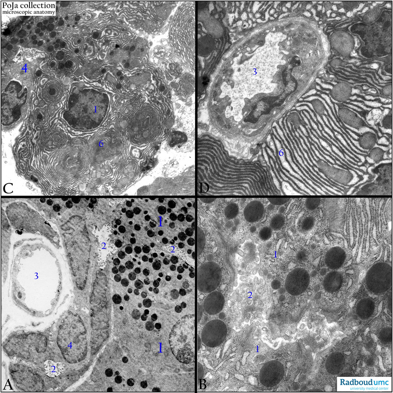
4.4.1 POJA-L-3544-3545+3546+3549
Title: Electron micrographs of secretory cells in pancreatic acinus (human)
Description: Electron microscopy.
(A): The basal side of an acinus is located near a blood capillary (3) providing a.o. the energy for the secretory activity of the acinar cells (1).
(2): Represents the lumen that continues into the intercalated ductal cells (4).
(B): Show details of the microvillar surface of the secretory area (2).
(C): The secretory cell can be filled up with ample whirled rough endoplasmic reticulum (RER) profiles (6) in whose cisterns (via the Golgi areas) the zymogen granules are produced. Electron-dense zymogen granules are located at the apical side of the cell above the nucleus.
(D) Shows a detail of a capillary (3) near the base of the secretory cell with extensive, dilated RER (6).
Keywords/Mesh: pancreas, exocrine, acinus, intercalated duct, electron microscopy, histology, POJA collection