2.3 POJA-L1036
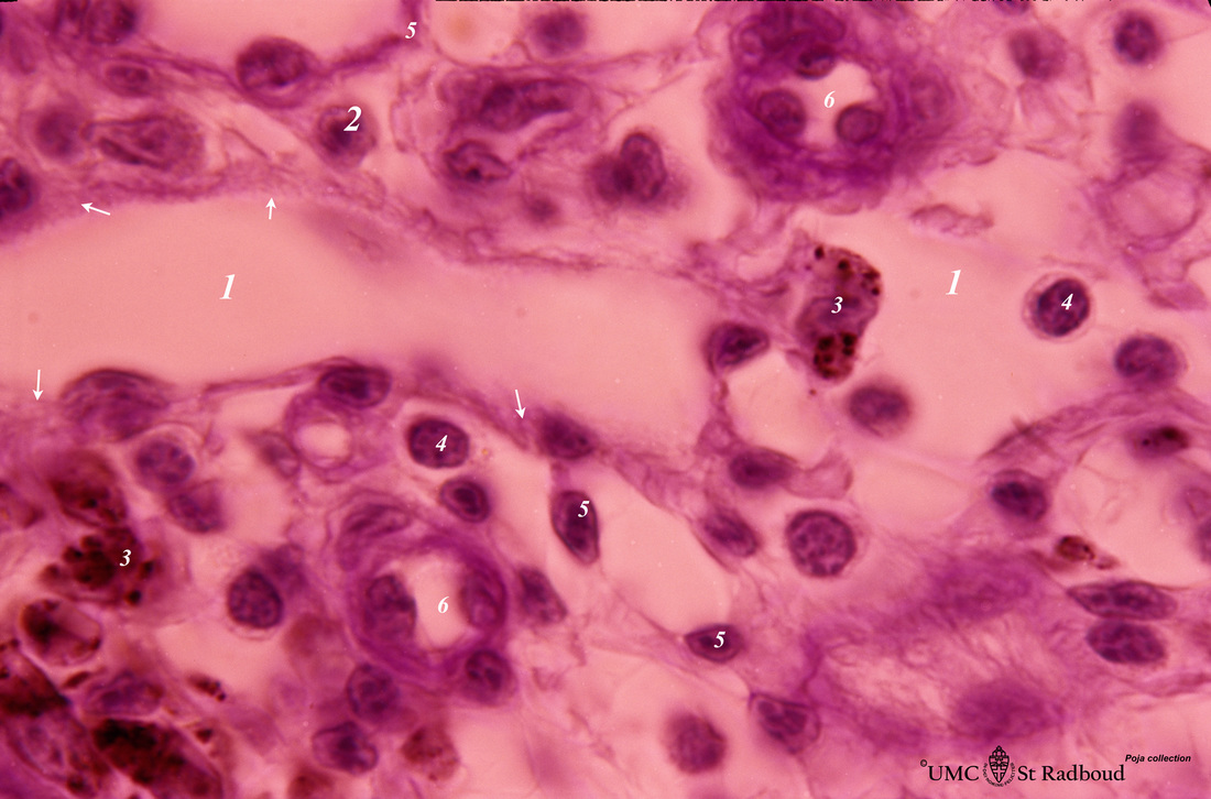
Title: Medullary cords and sinus in lymph node (mouse)
Description: Stain: Trichrome (Goldner).
Detail of medullary sinus (1) flanked by part of medullar cords (2). The sinus space (1) is lined by flattened littoral cells (foamed cytoplasm) (→) and dispersedly filled with lymphocytes and macrophages (brown hemo-pigment) (3). Within the medullary cords one finds reticular cells (5), lymphocytes and macrophages with phagocytised hemo-pigment (3). (6) ascending arterioles and arterial capillary within the cords.
Keywords/Mesh: lymphatic tissue, lymph node, medulla, medullar cord, medullar sinus, reticular tissue, histology, POJA collection

