2.4 POJA-L1087
Title: Appendix (‘gut-associated lymphatic tissue’ or GALT) (human)
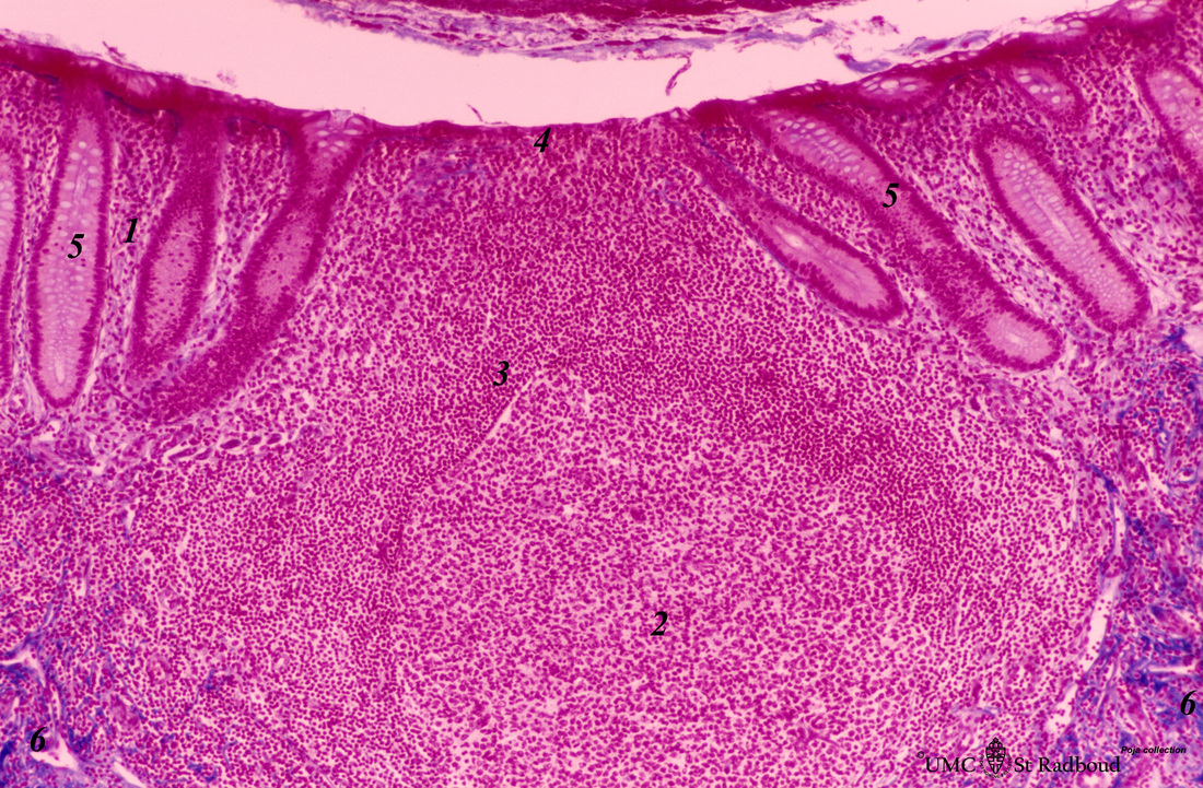
Description: Stain: Azan. Lymphatic nodule in vermiform appendix (see also Digestive System: Appendix)
A large amount of non-encapsulated diffuse lymphatic tissue or mucosa-associated lymphatic tissue (MALT) is located in the subepithelial lamina propria/submucosa of the appendix and called gut-associated lymphatic tissue (GALT).
A large nodule in the appendix extends through the proper lamina (1) and submucosa. The nodule is similar to that found in a lymph node with germinal centre (2) and darker-stained cap (crescent) (3) orientated towards the lumen of the gut showing a flattened dome area covered with discontinuous epithelial lining (4). Short crypts of Lieberkühn with columnar mucous epithelial cells (5) flank the dome area. (6) Connective tissue of submucosal layer. The follicles mostly contain B cell types while T cells are spread throughout the interfollicular areas. Plasma cells here produce largely dimeric IgA to be transported through the epithelial cells towards the lumen.
Keywords/Mesh: lymphatic tissue, MALT, GALT, appendix, lymphoepithelial tissue, reticular tissue, histology, POJA collection

