
13.1 POJA-L4517+4518+4519+4520+4521+4522 Myocardiocytes in fetal heart
13.1 POJA-L-4517+4518+4519+4520+4521+4522
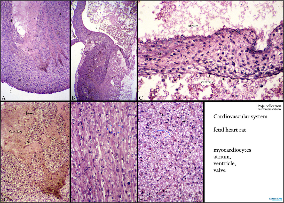
Title: Myocardiocytes in fetal heart
Description:
(A): Heart, Haematoxylin-azophloxine, fetus rat. Apex cordis (1) and epicardium (arrow 2) or visceral pericardium. The epicardium is a narrow layer of fibrocollagenous tissue containing elastic fibres, covered by flat mesothelial cells. The cavity of the ventricle (3) is filled with erythrocytes. The myocardial fibres (4) form an interconnecting network separated by loose fibrocollagenous tissue (endomysium). They are attached to the solid portion of the wall or myocardium (5). These muscular ridges are covered with endocardium (arrow 6) and are called trabeculae carneae in adulthood.
(B): Heart, Haematoxylin-azophloxine, fetus rat. Part of lumen ventricle (1), muscular ridges of trabeculae carneae (3) (rounded or irregular muscular columns projecting into the inner surface of the ventricle), at (4) cuspis of valva atrioventricularis (mitral valves) and atrium (2).
(C): Heart, cusp of atrioventricular valve (tricuspidalis) of right ventricle, Haematoxylin-azophloxine, fetus rat. At the atrium site the valve is lined by endocardium, a simple squamous epithelium overlying a delicate layer of loose connective tissue. (M) Indicates myofibroblastic cells at location of future smooth muscle cells. Atrium space above the valve is lined with temporarily isoprismatic mesothelial cells (arrows). Ventricle space below the valve is lined with flattened mesothelium (arrows).
(D): Heart muscle cells (myocardiocytes), Haematoxylin-azophloxine, fetus rat. The cavity of the ventricle is filled with blood cells and lined by endocardium (arrow 1). Muscular ridges or trabeculae carneae (2) which are attached to the solid portion of the myocardium (3).
(E): Longitudinal sectioned myocardiocytes, Haematoxylin-azophloxine, fetus rat.
(F): Transverse section of myocardiocytes, Haematoxylin-azophloxine, fetus rat. Numerous cells (= fibres) lack a central nucleus (blue encircled: white area’s), fine sarcoplasmic dots indicate cross-sectioned myofibrils. Arrows indicate blood capillaries between close-packed cardiocytes.
Keywords/Mesh: cardiovascular system, heart, fetus, ventricle, valve, myocardiocyte, cardiocyte, histology, POJA collection