4.1.1 POJA-La0168+L4022+4027+4028+4065+4030+4221+4064
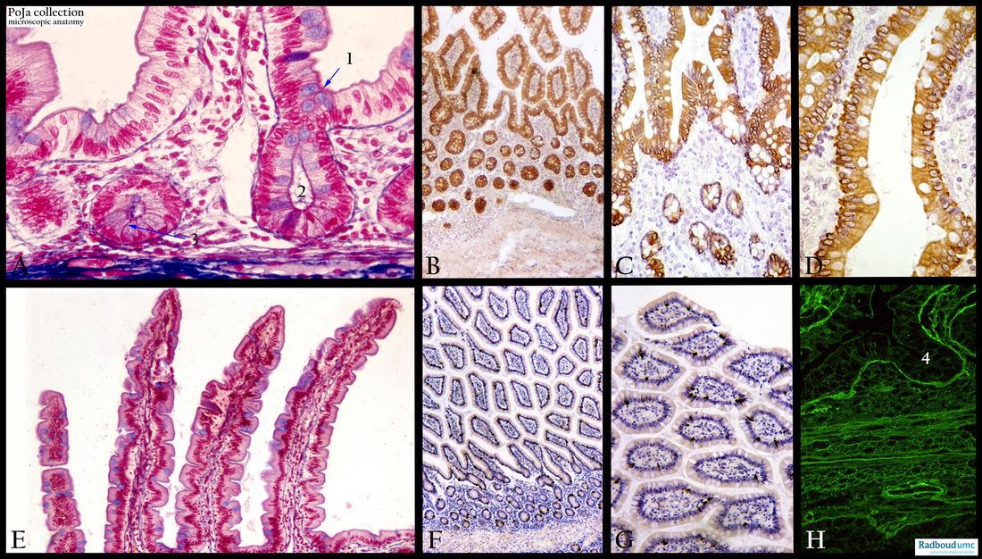
Title: Histochemical profile of jejunum (human, rat)
Description: Stain: (A, E) Azan. (B, C, D) Anti-polykeratin antibody immunoperoxidase staining with DAB and hematoxylin counterstaining. (F, G) Anti-keratin 7 antibody (OVTL12/30) immunoperoxidase staining with DAB and hematoxylin counterstaining.
(H) EV3C3 phage display antibody against heparin sulfate and immunofluorescence second antibody (rat).
(A, 1) The arrow points to the blue stained goblet cells present in the epithelial lining of villi (see also E) and crypts (2) in the jejunum.
(A, 3) Paneth cells are usually located at the bottom of the crypts.
(E) Several villi.
(B, C, D) The polykeratin antibodies stain all epithelial cell types including the goblet cells although they rather look unstained due to the filling up of the cytoplasm by the mucus secretory granules.
(F, G) anti-keratin-7 monoclonal antibodies also stain positive the jejunum cells. Remember that colon-epithelium is not positive for keratin-7.
(H) The immunofluorescence shows that heparan sulfate is strongly present in the connective tissue elements below the negative epithelial lining (4), such as the basal lamina, the fibroblast area and blood vessels.
Keywords/Mesh: jejunum, keratin-7, polykeratin, heparan sulfate, villi, crypts, goblet cells, immunofluorescence, hyistology, POJA collection

