3.6 POJA-L101A nr. 2
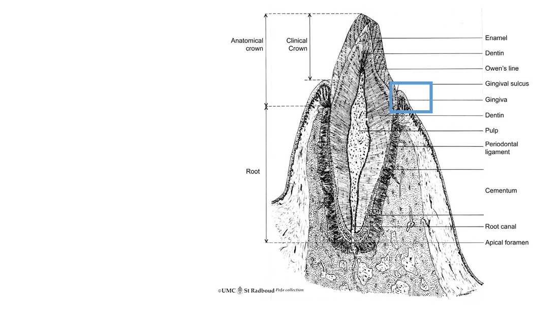
Title: Scheme survey tooth (longitudinal section)
Description:
Scheme canine tooth in osseous alveolus, human.
Keywords/Mesh: oral cavity, mouth, tooth, enamel, dentin, gingiva, gingival sulcus, junctional epithelium, pulp canal, alveolar bone, histology, POJA collection
Title: Scheme survey tooth (longitudinal section)
Description:
Scheme canine tooth in osseous alveolus, human.
Keywords/Mesh: oral cavity, mouth, tooth, enamel, dentin, gingiva, gingival sulcus, junctional epithelium, pulp canal, alveolar bone, histology, POJA collection
|
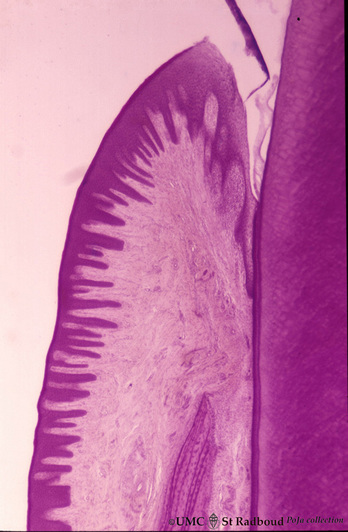
3.6 POJA-L17
Title: Tooth (‘free’ gingiva 4 of decalcified tooth) Description: Stain hematoxylin-eosin, human adult. Oral side (left) with stratified squamous epithelium, at the right side part of a tooth (dentin), at the bottom side part of the alveolar bone. The epithelium shows slight parakeratosis with many narrow deep papillae of the lamina propria (oral side). On the tooth-related side no papillae are present. On top the gingival crest with the gingival sulcus, the thinner sulcular epithelium lines the sulcus localized between the epithelium and enamel (dissolved). This epithelium becomes the specialized stratified junctional epithelium and is attached to the enamel. In these area generally chronic inflammatory cell infiltrations are common. |
|
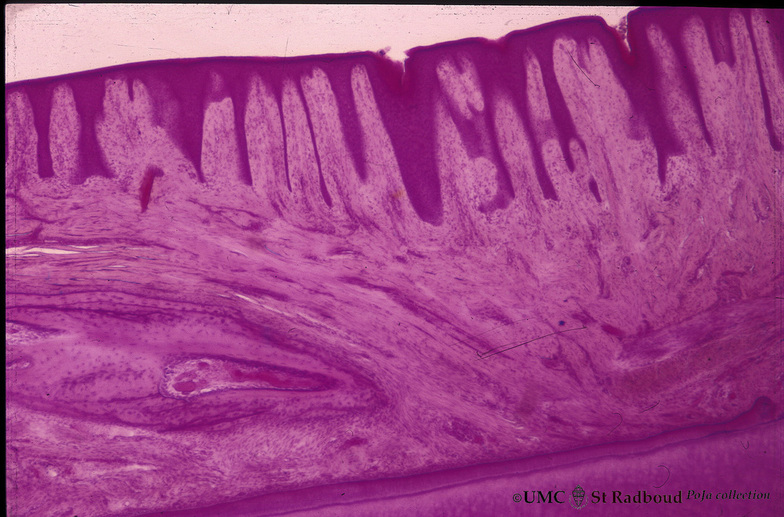
3.6 POJA-L66
Title: Gingiva (‘attached’ gingiva 1 of decalcified alveolar bone) Description: Stain hematoxylin-eosin, human adult. Stratified squamous epithelium with parakeratosis (reddish) and deep papillae of the lamina propria. At the bottom lamellar bone tissue (alveolar bone) with thickened periosteum (part of the periodontal ligament) followed by dentin. |


