6.1 POJA-La0180+La0181
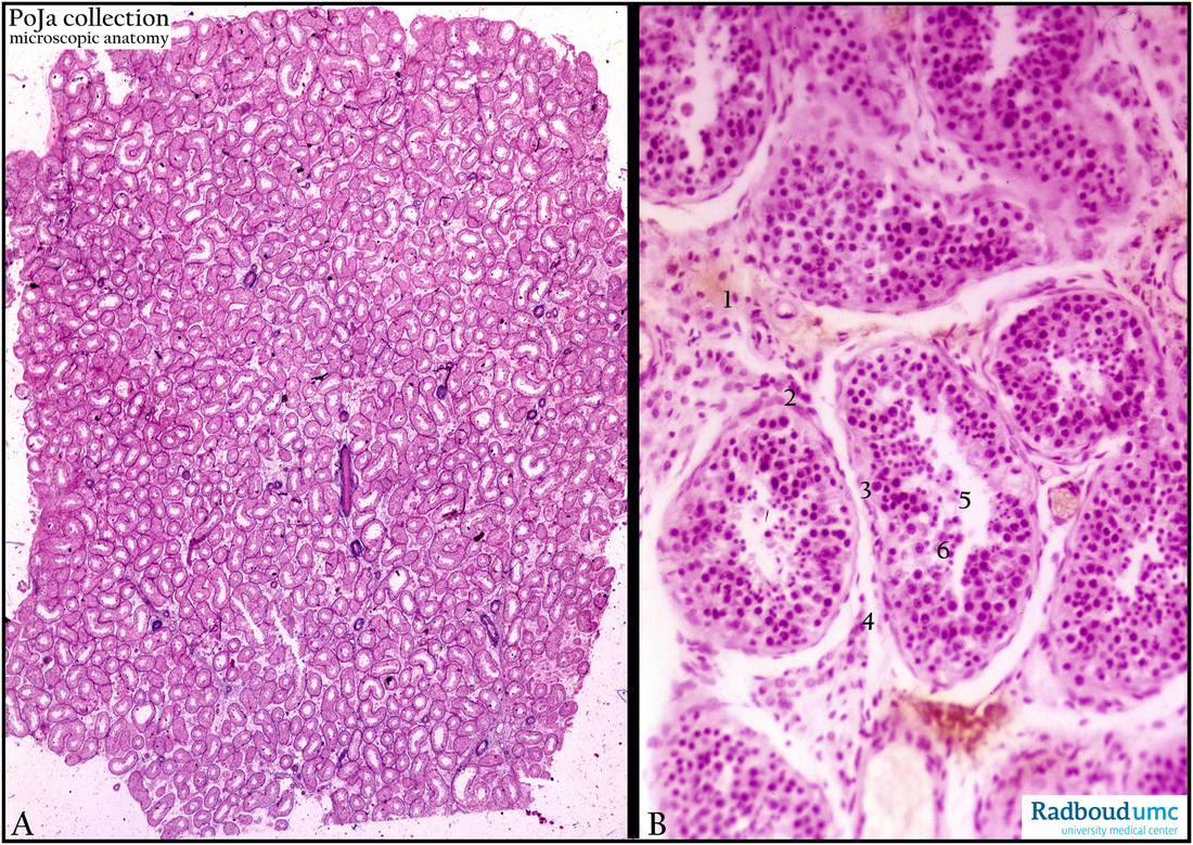
Title: Seminiferous tubules in testis
Description:
(A) Stain Azan, human. (B) Stain hematoxylin-eosin, human.
The survey (A) and the details in (B) show that tubules are coiled within the lobuli testis. The tunica albuginea is not displayed here. Between the tubules interstitial lymphoid infiltration is found. Leydig cells are here not observed. The seminiferous epithelium is in fact attached to the lamina propria limitans (2) consisting of myofibroblastic cell types. Supportive Sertoli cells (3) are recognizable by their prominent nucleolus in an otherwise pale looking nucleus. Their cell borders are hardly visible. The spermatogonia (4) are localized on the basal lamina with dark staining nuclei. From these cells arise differentiating spermatocytes (6) often remaining arranged in rows. The spermatids (5) are recognizable by their small condensed heads sticking into the Sertoli cells, and with their tails directed towards the lumen. Characteristic for the adult seminiferous tubules is the presence of all differentiation steps from spermatogonia towards spermatids in one section of the tubule.
Keywords/Mesh: testis, seminiferous tubule, spermatogonium, Sertoli cell, histology, POJA collection

