
11.5 POJA-L4454+3009+
4453+3011+3353.
Granular and agranular types cortex of the cerebrum
11.5 POJA-L4454+3009+4453+3011+3353
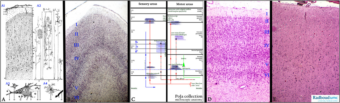
Title: Granular and agranular types cortex of the cerebrum
Description:
(A1-4): Scheme cerebral cortex, human (zoom in).
(1) Arachnoidea.
(2) Subarachnoideal space.
(3) Pia mater, followed by the cortex layers:
I. Molecular layer
II. External granular layer
III. External pyramidal layer
IV. Internal granular layer
V. Internal pyramidal layer
VI. Multiform layer.
(4) Medulla.
(A2): Signal scheme of the cerebrum: (5) Input signaling of the cerebrum. (6) Interneurons. (7) Output signaling.
(A3): Pyramid cell in motor cortex (stained for Nissl bodies).
(8) Dendrites. (11) Nissl body (RER and free ribosomes).
(9) Axon hillock. (12) Perikaryon of an interneuron (relay neuron).
(10) Axon. (13) Nuclei of glial cells.
(A4): Glial cell such as (14) astrocyte with perivascular endfeet. (15) Capillary.
(B): Stain cresyl violet, human. Visual cortex showing all layers numbered as shown in (A) and (C).
(C): Scheme displaying the difference between sensory part and motor part of the cortex, human:
The sensory cortex (granular type cortex) contain an extended internal granular layer (IV), while the motor cortex (agranular type cortex) has a well developed internal pyramidal layer (V) (large pyramidal cells of Betz).
(D): Stain cresyl violet, human. Visual cortex or striate cortex (granular type cortex).
The layers of the cortex are numbered as indicated in both schemes (A, C).
(E): Stain hematoxylin-eosin, human. Association cortex, compare with the granular type cortex in (D).
Keywords/Mesh: nervous tissue, cerebrum, visual cortex, associative cortex, motor cortex, neuron, interneuron, glia cell, axon, dendrite, histology, POJA collection