6.1 POJA-L4240+4250+2674
Title: Testis
Description:
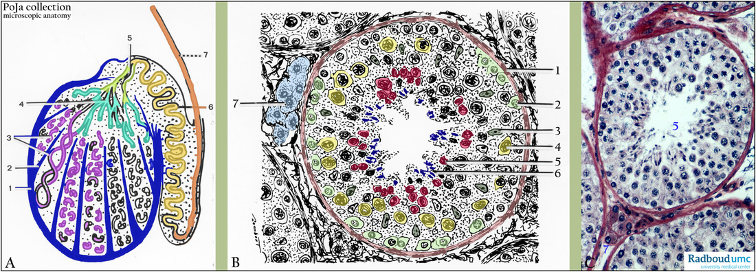
(A, B) schemes. (C) Stain iron hematoxylin-eosin, human.
(A): The general scheme of the testis with the epididymis in (A) shows that the testis is packed within a dense connective tissue layer,
the tunica albuginea (1), that further divides the testis into lobules via septa, (septulum testis) (2).
(3): Seminiferous tubules.
(4): Rete testis.
(5): Ductuli efferentes.
(6): Ductus epididymis.
(7): Ductus deferens.
(B): Tubulus seminiferous.
(1): Lamina propria limitans or boundary tissue containing also muscular cell types.
(2): Spermatogonia are attached to the basal lamina of (1).
They are large round cells with chromatin-rich nuclei.
(3): Nuclei of the supportive or nursing Sertoli cells.
(4): The primary spermatocytes (type I) are located above the spermatogonia.
(5): Secondary spermatocytes (type II).
(6): The spermatids with dense, small nuclei, fitting with their heads in cytoplasmic crypts of the Sertoli cells, are localized near the lumen. (7): Interstitial cells or the Leydig cells are localized between the seminiferous tubules. These cells produce androgens, testosterone and others. The Leydig cells are stimulated by pituitary hormone (LH). All cells together that rest on the basal lamina are collectively called germinal epithelium or seminiferous epithelium.
(C): See the numbers and arrows from (B) to (C).
Keywords/Mesh: testis, seminiferous tubule, seminiferous epithelium, sperm, Sertoli cell, Leydig cell, histology, POJA collection

