5.4.3 POJA-L2372+2432+2466+2465+2464+2463
Title: Intermediate tubules (IV) in the kidney
Description:
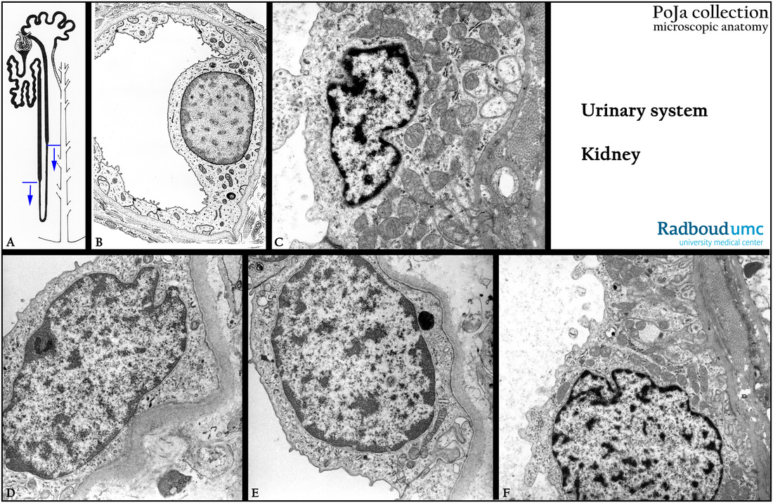
(A): Scheme nephron. The tubule between the two arrows is called intermediate tubule (IT) which comprises the ascending thin limb (ATL) and the descending thin limb (DTL) (thin loop of Henle) and appears much smaller and thinner than the other parts.
(B): Electron micrograph scheme of cross-section of a thin limb.
(C): Electron microscopy, descending thin limb (DTL), mitochondria and just a few basal infoldings still present.
(D, E): Electron microscopy, the thinnest part of the thin limb shows lining IT cells with scanty cytoplasm and organelles, no basal infoldings at all.
(F): Electron microscopy, ascending thin limb (ATL) with more cytoplasm, mitochondria, more and deeper basal infoldings.
Keywords/Mesh: urinary system, kidney, medulla, thin limb, loop of Henle, intermediate tubule, histology, electron microscopy, POJA collection

