12.1.4 POJA-L2562+2584+2564
Title: Retina IV
Description:
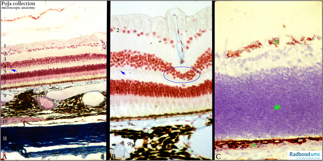
(A, B, detail): Retina, stain Azan, monkey. The retina is constructed with the following layers:
(1) Layer with optic nerve fibres and an inner limiting membrane.
(2) Ganglion cell layer.
(3) Inner plexiform layer.
(4) Inner nuclear layer.
(5) Outer plexiform layer; the blue arrows (in layer 5) points to row of synaptic contacts between photoreceptor axons and dendrites
of ganglion, amacrine, horizontal cells.
(6) Outer nuclear layer (nuclei of photoreceptor cells).
(7) Inner and outer segments of rods and cones.
(8) Pigmented epithelium (RPE).
(9) Choroid with blood vessels and large amounts of pigmented melanocytes.
(9a) Lamina choriocapillaris continued into the rest of the choroid (9b) with numerous blood vessels and heavily melanised melanocytes.
(10) Choroidal nerve, (branch of short posterior ciliary nerve).
(11) Sclera.
The area, marked with a blue ellips is a local indentation due to a large capillary on the point of branching from the nerve fibre layer through the ganglion cell layer into the inner plexiform layer.
(C): Immunoperoxidase staining with DAB and antibodies to collagen IV, 1d postnatal rat. In the postnatal rat the retinal layers (*) are not yet arranged in separate differentiated layers. The presence of the basal lamina collagen IV is only limited to the places where blood vessels and capillaries (12) are found in the future nerve fibre layer. The basement membrane and the close apposed choriocapillaris (9a) are distinct and regularly collagen IV reactive.
Keywords/Mesh: eye, retina, collagen IV, histology, POJA collection

