
16.1.3 POJA-L7145+7099 Endochondral ossification in foetus 8
16.1.3 POJA-L7145+7099 Endochondral ossification in foetus 8
16.1.3 POJA-L7145+7099 Endochondral ossification in foetus 8
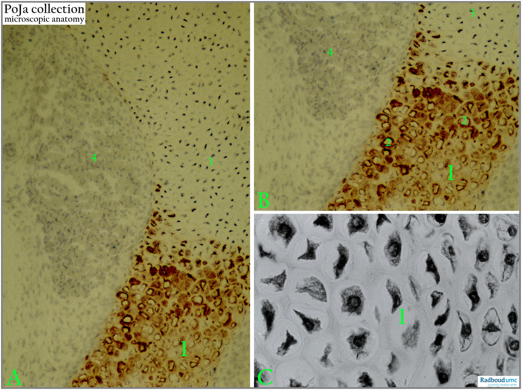
Title: Endochondral ossification in foetus 8
Description:
(A+B): Rat (1 day postnatally) labelled for proteoglycan 6S (found in the cartilage matrix).
(1): Pre-/hypertrophic zone with irregular dense labeling of the cytoplasms for proteoglycans.
(2): Unstained nuclei of hypertrophic chondrocytes.
(3): Epiphyse with smaller negative reacting chondrocytes in resting cartilage.
(4): Foetal strated muscle.
(C): (1): (Black and white): Human foetus, proliferation zone. At the right side smaller cells in the resting cartilage, at the left increased cytoplasm volumes of chondrocytes toward the direction of the hypertrophic zone.
Background:
The chondrocytes in the hypertrophic zone are induced by systemic and local soluble factors a.o. IGF-1 (insulin-like growth factor-1), transcription factors like RUNX2, TGF-B (transforming growth factor-beta) to undergo hypertrophy and ECM secretion. The major component of the ECM in the cartilage is formed by collagen fibres of type I, II, IX, X (especially type X is located around hypertrophic cells) embedded in a ground substance consisting of non-collagenous proteins such as proteoglycans (PG), glycoproteins e.g. chondronectin as well as growth factors and cytokines a.o. insulin-like growth factor (IGF’s), interleukins 1, 6. The PG’s are characteristic with their side chains of glycosaminoglycans such as chondroitin sulphate, keratan sulphate and hyaluronan combined with aggrecan. Especially the keratan sulphate and chondroitin sulphate chains of aggrecan bind to water. The matrix closely surrounding the cartilage cells contains strongly sulphatised glycosaminoglycans.
Reference
Role of Proteoglycans in Endochondral Ossification: Immunofluorescent Localization of Link Protein and Proteoglycan Monomer in Bovine Foetal Epiphyseal Growth Plate. The Journal of Cell Biology 92(2):249-60, 1982 A. ROBIN POOLE, I . PIDOUX, and L ROSENBERG
See also:
Keywords/Mesh: locomotor system, endochondral ossification, foetus, cartilage, chondrocyte, proteoglycan, histology, POJA collection
Title: Endochondral ossification in foetus 8
Description:
(A+B): Rat (1 day postnatally) labelled for proteoglycan 6S (found in the cartilage matrix).
(1): Pre-/hypertrophic zone with irregular dense labeling of the cytoplasms for proteoglycans.
(2): Unstained nuclei of hypertrophic chondrocytes.
(3): Epiphyse with smaller negative reacting chondrocytes in resting cartilage.
(4): Foetal strated muscle.
(C): (1): (Black and white): Human foetus, proliferation zone. At the right side smaller cells in the resting cartilage, at the left increased cytoplasm volumes of chondrocytes toward the direction of the hypertrophic zone.
Background:
The chondrocytes in the hypertrophic zone are induced by systemic and local soluble factors a.o. IGF-1 (insulin-like growth factor-1), transcription factors like RUNX2, TGF-B (transforming growth factor-beta) to undergo hypertrophy and ECM secretion. The major component of the ECM in the cartilage is formed by collagen fibres of type I, II, IX, X (especially type X is located around hypertrophic cells) embedded in a ground substance consisting of non-collagenous proteins such as proteoglycans (PG), glycoproteins e.g. chondronectin as well as growth factors and cytokines a.o. insulin-like growth factor (IGF’s), interleukins 1, 6. The PG’s are characteristic with their side chains of glycosaminoglycans such as chondroitin sulphate, keratan sulphate and hyaluronan combined with aggrecan. Especially the keratan sulphate and chondroitin sulphate chains of aggrecan bind to water. The matrix closely surrounding the cartilage cells contains strongly sulphatised glycosaminoglycans.
Reference
Role of Proteoglycans in Endochondral Ossification: Immunofluorescent Localization of Link Protein and Proteoglycan Monomer in Bovine Foetal Epiphyseal Growth Plate. The Journal of Cell Biology 92(2):249-60, 1982 A. ROBIN POOLE, I . PIDOUX, and L ROSENBERG
See also:
- 16.1.3 POJA-L7095+7098+7096 Endochondral ossification in foetus 1
- 16.1.3 POJA-L7104+7105+7100 Endochondral ossification in foetus 2
- 16.1.3 POJA-L7134 Endochondral ossification in foetus 3a
- 16.1.3 POJA-L3813 Endochondral ossification in foetus 3b
- 16.1.3 POJA-L7109+7108+7101+7097 Endochondral ossification in foetus 4
- 16.1.3 POJA-L7102+7103+7106+7107 Endochondral ossification in foetus 5
- 16.1.3 POJA-L7147+7111+7110 Endochondral ossification in foetus 6
- 16.1.3 POJA-L7116+7115+7123 Endochondral ossification in foetus 7
- 16.0.5 POJA-L7205 Bone: Introduction Bone formation-5 Long bones
Keywords/Mesh: locomotor system, endochondral ossification, foetus, cartilage, chondrocyte, proteoglycan, histology, POJA collection