
14.1 POJA-L6304B Electron micrograph of a motor endplate on skeletal muscle fibre (myoneural junction)
14.1 POJA-L6304B Electron micrograph of a motor endplate on skeletal muscle fibre (myoneural junction)
14.1 POJA-L6304B Electron micrograph of a motor endplate on skeletal muscle fibre (myoneural junction)
(By courtesy of L. Eshuis BSc Section Electron microscopy, Department of Pathology, Radboud university medical center, Nijmegen, the Netherlands)
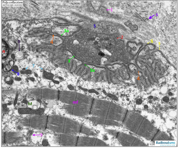
Title: Electron micrograph of a motor endplate on skeletal muscle fibre (myoneural junction)
Description:
The electron micrograph shows the motor endplate, i.e., an axon terminal ending on the sarcolemma of the muscle fibre (human).
The junction between the sarcolemma of a myofiber and an axon is characterized by strong folding of the sarcolemma (i.e., junctional folding) that forms primary clefts and secondary clefts. All these clefts are also described as subneural clefts or subsynaptic gutters. This subneural postsynaptic sarcolemma is also known as the sole plate (diameter about 50-60m). Between the free axon terminal stuffed with vesicles containing neurotransmitter and the sarcolemma lies the synaptic cleft (about 20-50nm).
(S): Schwann cell cytoplasm
(n): Nucleus of myofibre
(mf): Myofilaments of myofibre
(1): Projection of Schwann cell
(2): Axon terminal with abundant vesicles
(3): Synaptic cleft (20-50nm)
(3a): Primary cleft (or gutter)
(4): Secondary cleft (or gutter)
(5): Basal lamina of sarcolemma
(6): Collagen fibrils in interstitium
(7): Mitochondria (quite numerous in the region of the sole plate)
(8): Membranous whorls (remnants of mitochondria)
(9): Triad
See also:
Keywords/Mesh: locomotor system, skeletal muscle, striated muscle, synapse, motor endplate, myoneural junction, neuromuscular junction, electron microscopy, POJA collection
(By courtesy of L. Eshuis BSc Section Electron microscopy, Department of Pathology, Radboud university medical center, Nijmegen, the Netherlands)
Title: Electron micrograph of a motor endplate on skeletal muscle fibre (myoneural junction)
Description:
The electron micrograph shows the motor endplate, i.e., an axon terminal ending on the sarcolemma of the muscle fibre (human).
The junction between the sarcolemma of a myofiber and an axon is characterized by strong folding of the sarcolemma (i.e., junctional folding) that forms primary clefts and secondary clefts. All these clefts are also described as subneural clefts or subsynaptic gutters. This subneural postsynaptic sarcolemma is also known as the sole plate (diameter about 50-60m). Between the free axon terminal stuffed with vesicles containing neurotransmitter and the sarcolemma lies the synaptic cleft (about 20-50nm).
(S): Schwann cell cytoplasm
(n): Nucleus of myofibre
(mf): Myofilaments of myofibre
(1): Projection of Schwann cell
(2): Axon terminal with abundant vesicles
(3): Synaptic cleft (20-50nm)
(3a): Primary cleft (or gutter)
(4): Secondary cleft (or gutter)
(5): Basal lamina of sarcolemma
(6): Collagen fibrils in interstitium
(7): Mitochondria (quite numerous in the region of the sole plate)
(8): Membranous whorls (remnants of mitochondria)
(9): Triad
See also:
- 14.1 POJA-L6294-B Synapse scheme of a neuromuscular junction
Keywords/Mesh: locomotor system, skeletal muscle, striated muscle, synapse, motor endplate, myoneural junction, neuromuscular junction, electron microscopy, POJA collection