2.2 POJA-L978
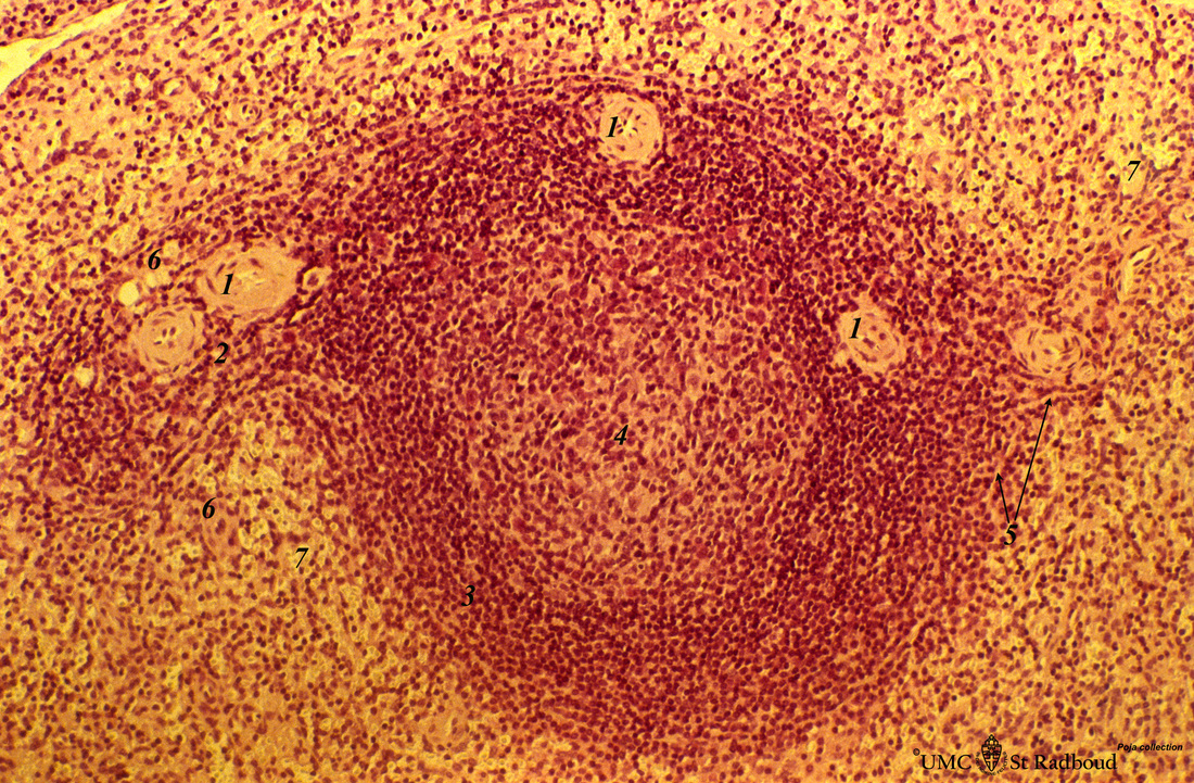
Title: Spleen with secondary lymphatic nodule (human)
Description: Stain: Hematoxylin & eosin.
The splenic follicle as part of the white pulp is arranged around the cross sectioned central artery (1).
The lymphatic sheath or PALS is composed of T cells (2).
The darker stained mantle zone of mainly naïve B lymphocytes (3) encompasses the lighter stained germinal centre (4) which contains reticular cells, B-memory lymphocytes and macrophages.
Here, the red pulp sinusoids directly surround the nodule (white pulp), and in this stage no marginal zone has yet developed.
At (5) thin penicillar arterioles and at (6) a macrophage-sheathed capillary.
(7): Sinuses.
Keywords/Mesh: lymphatic tissue, spleen, white pulp, histology, POJA collection

