16.1.3 POJA-L7085+7077+7078 Compact bone (cortical bone) with osteons and interstitial lamellae 3
16.1.3 POJA-L7085+7077+7078 Compact bone (cortical bone) with osteons and interstitial lamellae 3
Title: Compact bone (cortical bone) with osteons and interstial lamellae 3
Description:
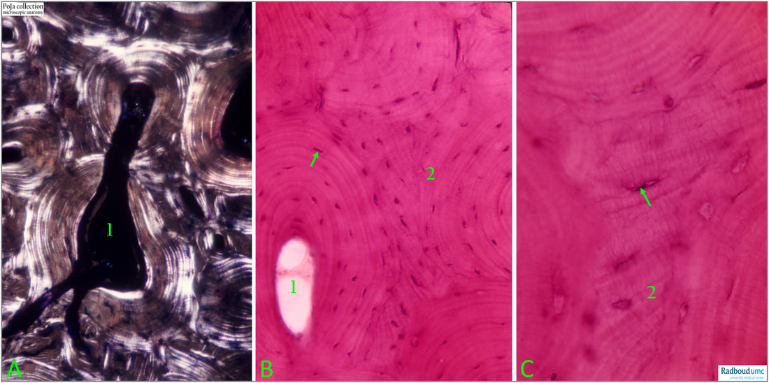
(A): Using polarisation microscopy the birefringence of the collagen fibres embedded in the matrix of osteons cause the alternate dark and light layers as structure of the osteon, with centrally the Haversian canal (1). Bone exhibits positive form birefringence dominated by and dependent upon the orientation of its collagen. Collagen is an anisotropic material. When collagen fibers are aligned perfectly transverse to the direction of the light propagation, there is a change in the refraction of light exiting the specimen, resulting in maximum brightness. When collagen fibers are aligned along the axis of light propagation, no refraction occurs and the specimen appears dark, or extinct. Collagen fibers oriented in other directions result in intermediate brightness values. With linearly polarised light a dark “Maltese Cross” effect is created against a bright background because the analyser and polariser filter transmission axes lie outside the vibration plane of light passing through lamellae at these positions.
(B and C): Haematoxylin-eosin stain of compact bone showing the osteocytes (arrow), Haversian canal (1) and interstitial lamellae (2).
Reference:
Circularly polarized light standards for investigations of collagen fiber orientation in bone. Timothy G. Bromage, Haviva M. Goldman, Shannon C. McFarlin, Johanna Warshaw, Alan Boyde, Christopher M. Riggs. https://doi.org/10.1002/ar.b.10031
See also:
Keywords/Mesh: locomotor system, bone, compact bone, osteon, Haversian canal, osteocytes, birefringe, collagen, Malthese cross effect, polarisation, histology, POJA collection
Title: Compact bone (cortical bone) with osteons and interstial lamellae 3
Description:
(A): Using polarisation microscopy the birefringence of the collagen fibres embedded in the matrix of osteons cause the alternate dark and light layers as structure of the osteon, with centrally the Haversian canal (1). Bone exhibits positive form birefringence dominated by and dependent upon the orientation of its collagen. Collagen is an anisotropic material. When collagen fibers are aligned perfectly transverse to the direction of the light propagation, there is a change in the refraction of light exiting the specimen, resulting in maximum brightness. When collagen fibers are aligned along the axis of light propagation, no refraction occurs and the specimen appears dark, or extinct. Collagen fibers oriented in other directions result in intermediate brightness values. With linearly polarised light a dark “Maltese Cross” effect is created against a bright background because the analyser and polariser filter transmission axes lie outside the vibration plane of light passing through lamellae at these positions.
(B and C): Haematoxylin-eosin stain of compact bone showing the osteocytes (arrow), Haversian canal (1) and interstitial lamellae (2).
Reference:
Circularly polarized light standards for investigations of collagen fiber orientation in bone. Timothy G. Bromage, Haviva M. Goldman, Shannon C. McFarlin, Johanna Warshaw, Alan Boyde, Christopher M. Riggs. https://doi.org/10.1002/ar.b.10031
See also:
- 16.1.3 POJA-L7091+7073+7075+7094 Compact bone (cortical bone) with osteons 1
- 16.1.3 POJA-L7079+7080+7082+7081 Compact bone (cortical bone) with osteons 2
- 16.1.3 POJA-L7083+7084 Blood vessels in compact bone (cortical bone) 4
- 16.1.3 POJA-L7114+7112+7093+7092 Remodeling of compact bone (cortical bone) 5
- 16.1.3 POJA-L7087+7088 Partially burned human bone specimen
- 16.1.3 POJA-L7090+7076 Bone: osteons in polarisation microscope
Keywords/Mesh: locomotor system, bone, compact bone, osteon, Haversian canal, osteocytes, birefringe, collagen, Malthese cross effect, polarisation, histology, POJA collection

