
3.6 POJA-L102 nr. 2. Premolar permanent tooth
3.6 POJA-L102 nr. 2
Title: Premolar permanent tooth
Description:
Low magnification of labiolingual section, stain hematoxylin-eosin, human adult. From top to bottom:
Keywords/Mesh: oral cavity, mouth, tooth, dentin, pulp canal, gingiva, gingival pocket, junctional epithelium, acellular cementum, histology, POJA collection
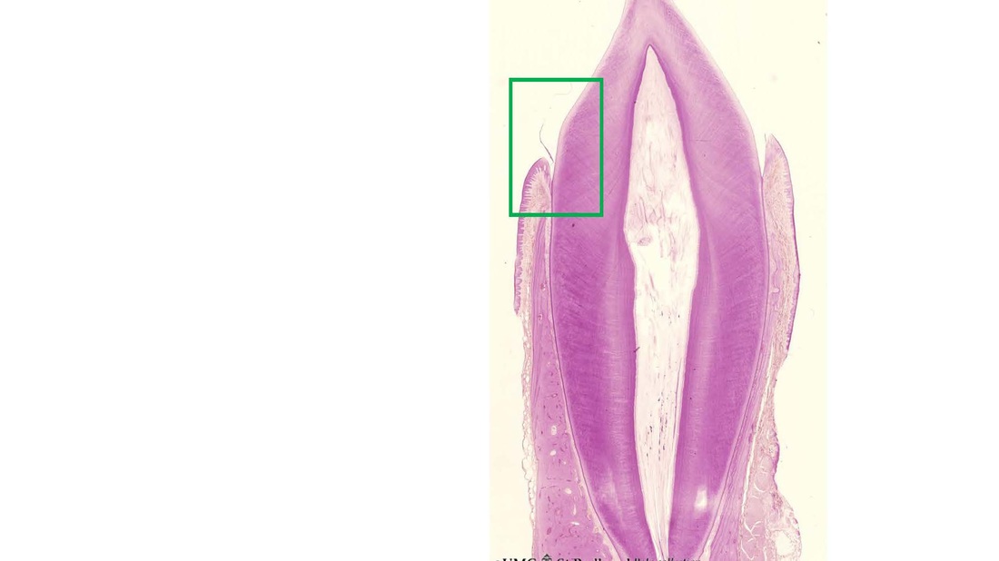
Title: Premolar permanent tooth
Description:
Low magnification of labiolingual section, stain hematoxylin-eosin, human adult. From top to bottom:
- Crown region with enamel-dentin boundary (there is no enamel present in decalcified specimens).
- Dentin (blue-pink) enwraps the whole pulp chamber (light) and here the lines of dentinal tubules appear S-shaped.
- Neck region at the attachment of the gingiva to dentin (left and right); from the neck region till the bottom a thin layer of cement fixed by the periodontal ligament to the alveolar bone is present (left and right side).
- Dentin root region as the main part of the tooth where dentinal tubules are more linear.
- At the bottom pulp canal and apical foramen (passage for alveolar nerve and vessels).
Keywords/Mesh: oral cavity, mouth, tooth, dentin, pulp canal, gingiva, gingival pocket, junctional epithelium, acellular cementum, histology, POJA collection
3.6 POJA-L21.
Tooth (‘free’ gingiva 1 of decalcified tooth)
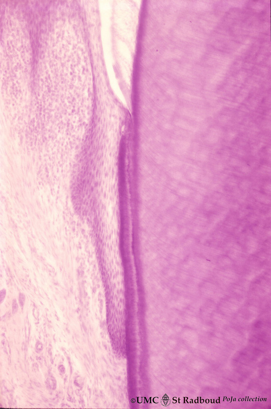
3.6 POJA-L21
Title: Tooth (‘free’ gingiva 1 of decalcified tooth) Description: Stain hematoxylin-eosin, human adult. On top part of the gingival sulcus, at the left stratified junctional epithelium normally attached to the surface of the enamel (dissolved due to decalcification). Top left shows foci of chronic inflammatory cell infiltration, left side faint stained bundle of fibers (periodontal ligament). In this case the junctional epithelium runs downwards still attached to the layer of acellular cementum. At the right dentin with faint striation due to presence of dentinal tubules. |

3.6 POJA-L20.
Tooth (‘free’ gingiva 2 of decalcified tooth)
3.6 POJA-L20
Title: Tooth (‘free’ gingiva 2 of decalcified tooth) Description: Stain hematoxylin-eosin, human adult. On top the gingival sulcus; at the left stratified junctional epithelium (normally attached to the surface of the enamel). Below the epithelium a focus of chronic inflammatory cell infiltration, followed by a bundle of fibers of the periodontal ligament. Continuous with the junctional epithelium a layer of acellular cementum. At the right dentin with faint striation due to presence of dentinal tubules. |