14.6.1 POJA-L6216+6192+6193+6302+6303 Mitochondrial myopathy II: Ultrastructure of mitochondria (human)
14.6.1 POJA-L6216+6192+6193+6302+6303 Mitochondrial myopathy II: Ultrastructure of mitochondria (human)
(B, C, D: by courtesy of Mrs.M. Heyman-de Bruyn, the former Department of Submicroscopic Morphology, Radboud university medical center, Nijmegen, The Netherlands)
Title: Mitochondrial myopathy II: Ultrastructure of mitochondria (human)
Description:
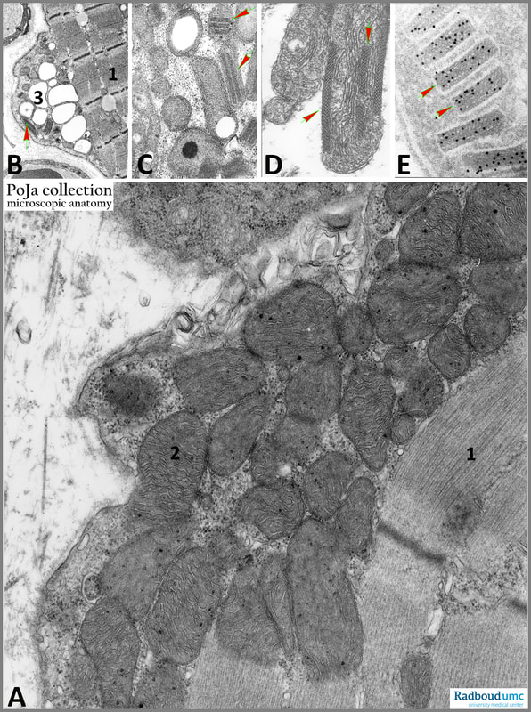
(A): Normal skeletal muscle with normal accumulation of mitochondria at the periphery of the fibre. The mitochondria contain well developed cristae and small dense mitochondrial granules are present.
(1): Bundles of myofilaments with A- and I-banding.
(2): Mitochondrion.
(B): Mitochondrial myopathy with vacuolised mitochondria (3) and crystalline deposits (arrow) in the so-called ‘ragged-red’ fibres (in light microscopy).
(C): Abnormal mitochondria and crystalline deposits (arrows). Note electron-dense lipid droplet as well as electron-lucent lipid droplet.
(D): Abnormal mitochondria (increased number of longitudinal arranged cristae) and crystalline deposits (arrow) in several cristae.
(E): ‘Ragged-red’ fibre in immuno-electron microscopy: part of a mitochondrion with crystalline inclusions labelled with a gold-conjugated antibody to creatine kinase indicating the presence of this enzyme as a constituent of these crystalline inclusions.
Background:
All of the mitochondrial myopathies are characterised by a disturbed mitochondrial metabolism and the presence of ‘ragged-red’ fibres in muscle biopsy specimens. Main problems as defects in oxidative phosphorylation, increased lactate production (lactic acidosis), free radical production and phosphocreatine depletion occur predominantly in skeletal muscles.
As already mentioned in 14.6.1 POJA-L6190B+6188+6189 Mitochondrial myopathy I , the cause of the ‘ragged’ appearance is the accumulation of abnormal mitochondria (as confirmed in the above figures B/C/D/E) showing highly ordered crystal-like inclusions of mitochondrial creatine kinase (Mi-CK) within the intermembrane spaces. In a normal mitochondrion Mi-CK is localised along the entire inner mitochondrial membrane as well as at the periphery where inner and outer mitochondrial membranes are in close apposition.
Mi-CK facilitates metabolic channelling of phosphates at the mitochondrial contact sites. Here the enzyme is functionally coupled to the ADP/ATP translocator of the inner and to porin of the outer mitochondrial membrane. Mi-CK provides phosphocreatine as a source of high-energy phosphate to sarcoplasmic creatine kinases that are coupled to energy-consuming processes. Afterwards creatine diffuses back to the mitochondria completing the phosphocreatine shuttle.
It is proposed that the Mi-CK inclusions associated with the ‘ragged-red’ fibres are the results of overexpression and further accumulation of Mi-CK induced by metabolic stress and the depletion of phosphocreatine. Subsequently after a certain threshold concentration of Mi-CK is reached, the result is a spontaneous crystallisation of the enzyme leading to pathological accumulation of Mi-CK crystals. Purified Mi-CK forms filaments or sheet-like structures in vitro and Mi-CK appears very stable in its crystalline form and remains enzymatically active.
See also:
Keywords/Mesh: locomotor system, skeletal muscle, striated muscle, neuromuscular disease, myopathy, mitochondrial myopathy, mitochondrion, ragged-red fibre, creatine kinase, crystallin inclusion, electron microscopy, pathology, POJA collection
Title: Mitochondrial myopathy II: Ultrastructure of mitochondria (human)
Description:
(A): Normal skeletal muscle with normal accumulation of mitochondria at the periphery of the fibre. The mitochondria contain well developed cristae and small dense mitochondrial granules are present.
(1): Bundles of myofilaments with A- and I-banding.
(2): Mitochondrion.
(B): Mitochondrial myopathy with vacuolised mitochondria (3) and crystalline deposits (arrow) in the so-called ‘ragged-red’ fibres (in light microscopy).
(C): Abnormal mitochondria and crystalline deposits (arrows). Note electron-dense lipid droplet as well as electron-lucent lipid droplet.
(D): Abnormal mitochondria (increased number of longitudinal arranged cristae) and crystalline deposits (arrow) in several cristae.
(E): ‘Ragged-red’ fibre in immuno-electron microscopy: part of a mitochondrion with crystalline inclusions labelled with a gold-conjugated antibody to creatine kinase indicating the presence of this enzyme as a constituent of these crystalline inclusions.
Background:
All of the mitochondrial myopathies are characterised by a disturbed mitochondrial metabolism and the presence of ‘ragged-red’ fibres in muscle biopsy specimens. Main problems as defects in oxidative phosphorylation, increased lactate production (lactic acidosis), free radical production and phosphocreatine depletion occur predominantly in skeletal muscles.
As already mentioned in 14.6.1 POJA-L6190B+6188+6189 Mitochondrial myopathy I , the cause of the ‘ragged’ appearance is the accumulation of abnormal mitochondria (as confirmed in the above figures B/C/D/E) showing highly ordered crystal-like inclusions of mitochondrial creatine kinase (Mi-CK) within the intermembrane spaces. In a normal mitochondrion Mi-CK is localised along the entire inner mitochondrial membrane as well as at the periphery where inner and outer mitochondrial membranes are in close apposition.
Mi-CK facilitates metabolic channelling of phosphates at the mitochondrial contact sites. Here the enzyme is functionally coupled to the ADP/ATP translocator of the inner and to porin of the outer mitochondrial membrane. Mi-CK provides phosphocreatine as a source of high-energy phosphate to sarcoplasmic creatine kinases that are coupled to energy-consuming processes. Afterwards creatine diffuses back to the mitochondria completing the phosphocreatine shuttle.
It is proposed that the Mi-CK inclusions associated with the ‘ragged-red’ fibres are the results of overexpression and further accumulation of Mi-CK induced by metabolic stress and the depletion of phosphocreatine. Subsequently after a certain threshold concentration of Mi-CK is reached, the result is a spontaneous crystallisation of the enzyme leading to pathological accumulation of Mi-CK crystals. Purified Mi-CK forms filaments or sheet-like structures in vitro and Mi-CK appears very stable in its crystalline form and remains enzymatically active.
See also:
- https://emedicine.medscape.com/article/1869808-overview#a7 Mitochondrial Disorder: Kearns-Sayre Syndrome Tsang, S.H., Aycinena, A.R.P., Sharma T. DOI: 10.1007/978-3-319-95046-4 30
- Mitochondrial creatine kinase: A major constituent of pathological inclusions seen in mitochondrial myopathies Stadhouders, A.M., Jap, P.H.K., Winkler, H-P., Eppenberger H.M., Wallimann, T. Proc Natl Acad Sci USA 91, 5089-5093, 1994) (https://www.pnas.org/content/91/11/5089
Keywords/Mesh: locomotor system, skeletal muscle, striated muscle, neuromuscular disease, myopathy, mitochondrial myopathy, mitochondrion, ragged-red fibre, creatine kinase, crystallin inclusion, electron microscopy, pathology, POJA collection

