2.2 POJA–L980+979B
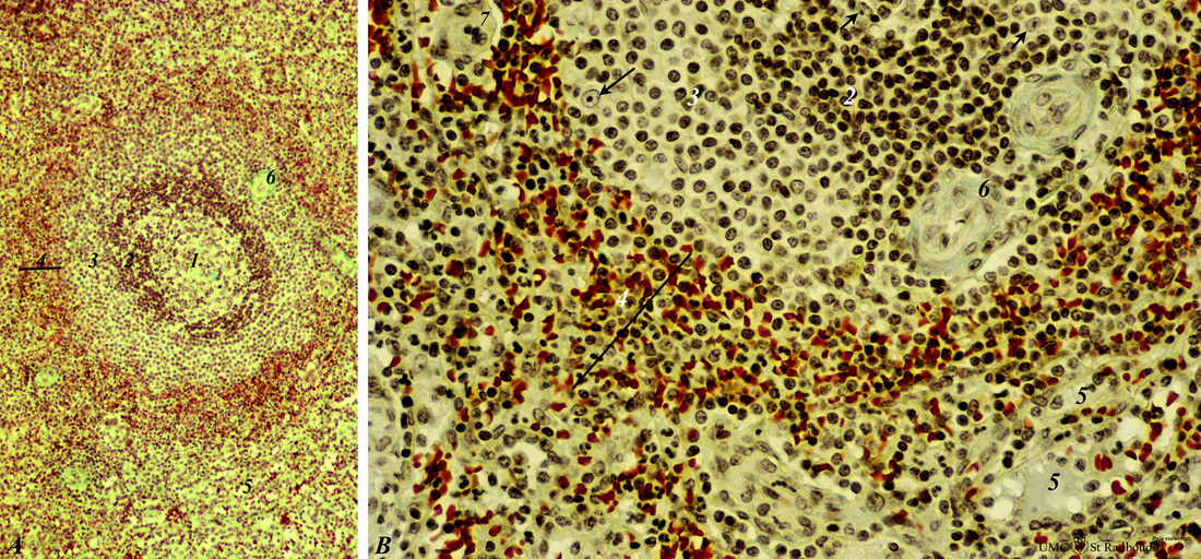
Title: Zone of marginal sinuses in the spleen (human)
Description: Stain: Trichrome (Goldner).
(A): Survey of a follicle in the spleen.
(B): A higher magnification of a similar section shows part of a lymph nodule (follicle, white pulp) with cross-sections (6) of the central artery.
(1): Germinal centre.
(2): Mantle layer.
(3): Marginal zone.
(4): Zone with red pulp sinuses apposing the marginal zone at the border between red pulp and white pulp.
(5): Venous sinuses. (→) indicate dendritic cells (antigen-presenting cells, APC) showing a light nucleus and a distinct nucleolus.
(7): A macrophage-sheathed capillary.
The sinusoids in (4) are filled with red blood cells (red).
Keywords/Mesh: lymphatic tissue, spleen, white pulp, histology, POJA collection

