4.1.1 POJA-L4226+La-0171
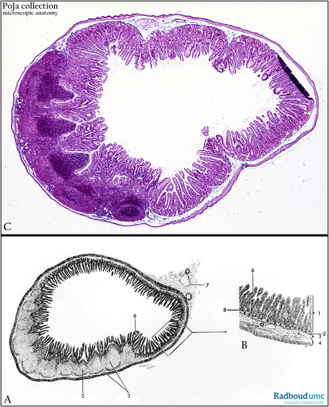
Title: Peyer’s patches in ileum (human, dog)
Description: Stain: (A, B) Schemes; (C) Trichrome-Goldner, dog.
The Patches of Peyer are aggregates of lymph nodes in the wall of the ileum, located in the submucosa and mucosa area. This immune system is crucial in the immunological defense of the digestive tract. The usual composition is T- and B-lymphocytes, macrophages, and antigen presenting cells among which a peculiar cell type, M-cell, is present. The Peyer’s patches are located at one site of the digestive tube. The surface enlargement in the ileum is much less than in the jejunum, i.e. fewer and lower plicae circulares, the villi are shorter and scarcer as well.
(A, B) Scheme.
(1) Tunica mucosa.
(2) Lamina muscularis mucosae.
(3) Tela submucosa.
(4) Tunica muscularis.
(5) Folliculi lymphatici aggregate (plaques of Peyer) in tela submucosa.
(6) Villus.
(7) Mesenterium.
(8) Crypt.
Keywords/Mesh: ileum, Peyer’s patches, lymphoid follicles, histology, POJA collection

