
13.1 POJA-L4737+4545
Macroscopy heart and mitral valve (human)
13.1 POJA-L4737+4545
Title: Macroscopy heart and mitral valve (human)
Description:
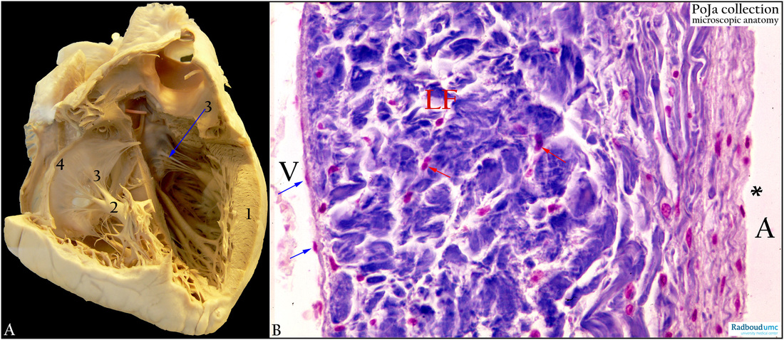
(A): Macroscopy of heart, human. Thick ventricle wall (1), papillary muscle (2), chordae tendineae (arrow 3), atrioventricular valve leaflet (4).
(B): Heart, ventricle, Azan, human. Close to the outer margin of the atrioventricular valve (mitral valve). The valve is an endocardial fold composed of a kind of fibroelastic connective tissue without any vascularization. The side of the mitral leaflet facing the atrium (A) is marked by (*). The side of the mitral valve facing the ventricle (V) has a lamina fibrosa (LF) with dense arranged connective tissue. The fibroblasts (red arrows) resemble the ‘wing’ cells of the tendons. (blue arrows) Endothelium of the endocard.
Keywords/Mesh: cardiovascular system, heart, atrium, ventricle, mitral valve, papillary muscle, chorda tendinae, histology, macroscopy, POJA collection
Title: Macroscopy heart and mitral valve (human)
Description:
(A): Macroscopy of heart, human. Thick ventricle wall (1), papillary muscle (2), chordae tendineae (arrow 3), atrioventricular valve leaflet (4).
(B): Heart, ventricle, Azan, human. Close to the outer margin of the atrioventricular valve (mitral valve). The valve is an endocardial fold composed of a kind of fibroelastic connective tissue without any vascularization. The side of the mitral leaflet facing the atrium (A) is marked by (*). The side of the mitral valve facing the ventricle (V) has a lamina fibrosa (LF) with dense arranged connective tissue. The fibroblasts (red arrows) resemble the ‘wing’ cells of the tendons. (blue arrows) Endothelium of the endocard.
Keywords/Mesh: cardiovascular system, heart, atrium, ventricle, mitral valve, papillary muscle, chorda tendinae, histology, macroscopy, POJA collection