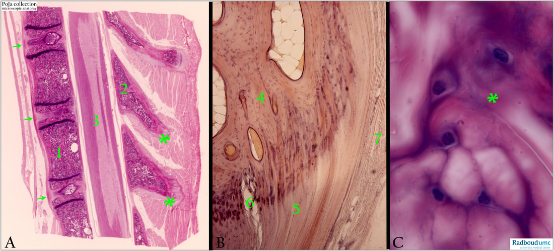
15.3 POJA-L7036A+7037+7038 Fibrocartilage
15.3 POJA-L7036A+7037+7038 Fibrocartilage
Title: Fibrocartilage
Description:
(A-C): Human.
(A): Survey of longitudinal section vertebral column with spinal cord, Haematoxylin-eosin stained.
(B): Longitudinal section of bone, Haematoxylin-azophloxine stained.
(C): Detail of fibrocartilage, Haematoxylin-eosin stained.
(1): Bony vertebral body.
(2): Bony spinous process.
(3): Spinal cord.
(4): Lamellar bone
(5): Tendon
(6): Zone of fibrocartilage in abrupt transition into tendon.
(7): Dermis of skin.
(* in A , C): connection of the spinous processes with the fasciae thoracolumbalis where fibrocartilage is present. In (C) small fibrochondrocytes as well as larger, swollen ones (nuclei are not always sectioned).
(Arrows in A): Loose anterior longitudinal ligament close to anulus fibrosus of the intervertebral disc. At the level of the arrows the three intervertebral discs are shown with centrally the nucleus pulposus
(for detail see below 16.0 POJA L7138+7139+7140, image A).
Background:
Hard and non-elastic fibrocartilage (FC) is the strongest type of cartilage and is resilient to tensile forces. There is no perichondrium, it contains an abundancy of type I and a low quantity of type II collagen, the so-called fibrochondrocytes are found solitary of as axially arranged isogenous cell groups.
On base of their presence and functions they are categorised as:
(a): Connecting FC (e.g. invertebral disc) as a pad against stresses.
(b): Intra-articular FC (e.g. meniscus) as thrust-pads and to prevent joint instability.
(c): Circumferential FC (e.g. acetabular labrum) for protection of joint margins.
(d): Stratiform FC (e.g. tendon of peroneus longus) as a thin layer over the bone to minimize friction where tendons may glide.
The FC intervertebral discs join adjacent vertebral bodies where they provide shock absorption and facilitate mobility of the spine.
Each FC disc has three components:
(1): The nucleus pulposus i.e. the gelatinous inner core of the intervertebral discs;
(2): The anulus fibrosus i.e. a fibrous capsule that surrounds the nucleus pulposus and consists of concentric lamellae of collagen fibres;
(3): The superior and inferior cartilaginous end plates situated at the articular surfaces of the intervertebral disc and the adjacent vertebrae.
Intervertebral disc degeneration is characterised by loss of proteoglycans and hydration resulting in a more fibrous disc that is unable to distribute stresses equally (adapted from Mandle et al. 2010).
See also:
Key words/Mesh: locomotor system, cartilage, bone, vertebral body, fibrocartilage, invertebral disc, anulus fibrosus, histology, POJA collection
Title: Fibrocartilage
Description:
(A-C): Human.
(A): Survey of longitudinal section vertebral column with spinal cord, Haematoxylin-eosin stained.
(B): Longitudinal section of bone, Haematoxylin-azophloxine stained.
(C): Detail of fibrocartilage, Haematoxylin-eosin stained.
(1): Bony vertebral body.
(2): Bony spinous process.
(3): Spinal cord.
(4): Lamellar bone
(5): Tendon
(6): Zone of fibrocartilage in abrupt transition into tendon.
(7): Dermis of skin.
(* in A , C): connection of the spinous processes with the fasciae thoracolumbalis where fibrocartilage is present. In (C) small fibrochondrocytes as well as larger, swollen ones (nuclei are not always sectioned).
(Arrows in A): Loose anterior longitudinal ligament close to anulus fibrosus of the intervertebral disc. At the level of the arrows the three intervertebral discs are shown with centrally the nucleus pulposus
(for detail see below 16.0 POJA L7138+7139+7140, image A).
Background:
Hard and non-elastic fibrocartilage (FC) is the strongest type of cartilage and is resilient to tensile forces. There is no perichondrium, it contains an abundancy of type I and a low quantity of type II collagen, the so-called fibrochondrocytes are found solitary of as axially arranged isogenous cell groups.
On base of their presence and functions they are categorised as:
(a): Connecting FC (e.g. invertebral disc) as a pad against stresses.
(b): Intra-articular FC (e.g. meniscus) as thrust-pads and to prevent joint instability.
(c): Circumferential FC (e.g. acetabular labrum) for protection of joint margins.
(d): Stratiform FC (e.g. tendon of peroneus longus) as a thin layer over the bone to minimize friction where tendons may glide.
The FC intervertebral discs join adjacent vertebral bodies where they provide shock absorption and facilitate mobility of the spine.
Each FC disc has three components:
(1): The nucleus pulposus i.e. the gelatinous inner core of the intervertebral discs;
(2): The anulus fibrosus i.e. a fibrous capsule that surrounds the nucleus pulposus and consists of concentric lamellae of collagen fibres;
(3): The superior and inferior cartilaginous end plates situated at the articular surfaces of the intervertebral disc and the adjacent vertebrae.
Intervertebral disc degeneration is characterised by loss of proteoglycans and hydration resulting in a more fibrous disc that is unable to distribute stresses equally (adapted from Mandle et al. 2010).
See also:
- 16.0 POJA-L7138+7139+7140 Section through two vertebral bodies with an intervertebral disc
- The normal joint P. Mandle, S. Benis, L. Patonay, P.V. Balint in Essential Applictions of musculoskeletal ultrasoumd in rheumatology Chapter 5, 51-64, 2010
Key words/Mesh: locomotor system, cartilage, bone, vertebral body, fibrocartilage, invertebral disc, anulus fibrosus, histology, POJA collection

