
4.2.1 POJA-L3647+3648+3649. Erythropoietic islets in fetal liver (rat)
4.2.1 POJA-L-3647+3648+3649
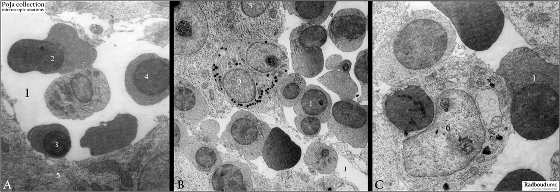
Title: Erythropoietic islets in fetal liver (rat)
Description: Electron micrographs of liver sinusoid with clusters of differentiating blood cells.
(A) Sinusoid space (1) with normoblast stages (2, 3, and 4). Liver parenchymal cell (5).
(B) Liver sinusoid with many normoblast cell types (1). (2) Myelocyte, the cell has small electron dense granules, well developed Golgi and cisterns. (5) Liver cell.
(C) Liver sinusoid with several normoblast cell types (1). Cell (6) is a reticular cell with remnants of a phagocytized normoblast nucleus in a large phagolysosome. (By courtesy of the former Dr. G. Zaki, New Jersey, U.S.A.)
Keywords/Mesh: liver cell, hematopoiesis, fetus, electron microscopy, histology, POJA collection