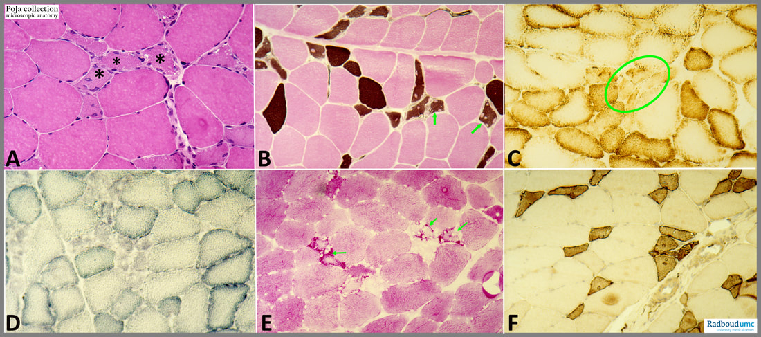
14.6 POJA-L6282+6280+6267+6265+6278+6272 Histochemical Profile of Rhabdomyolysis I (human)
..14.6 POJA-L6282+6280+6267+6265+6278+6272 Histochemical Profile of Rhabdomyolysis I (human)
(By courtesy of P. Poels MD, PhD , neurologist, (Dept. Neurology Radboud university medical center, Nijmegen, The Netherlands H. ter Laak PhD Section Neuropathology, retired staff member Department of Pathology, Radboud university medical center, Nijmegen, The Netherlands)
Title: Histochemical Profile of Rhabdomyolysis I (human)
Description:
(A): Haematoxylin-eosin staining shows myofibres of various sizes and quite a number of small muscle fibres, some contain swollen central (or internal) nuclei. Unstained vacuoles are also present at the periphery of the fibres. Asterisk indicates the smaller muscle fibres.
(B): ATPase staining, (pH4.2) shows strongly positive type I fibres (brown), type II fibres are negative (pink) and the smallest fibres that stain intermediary brown are referred as type regenerating fibres (type IIC fibres). Arrows point to these small vacuolised regenerating fibres.
(C): Cytochrome C oxidase staining. Note that the small muscle fibres (oval circle) show a low enzyme activity.
(D): The muscle fibres with low SDH activity are mostly small.
(E): PAS staining. Compared to the diffuse staining in type I and II fibres, regenerating fibres (arrows) contain distinct glycogen with the periodic acid Schiff (PAS) reaction. Small peripheral vacuoles are PAS negative and appear empty.
(F): Leu19 immunostaining. Leu19-Ag is expressed on muscle-specific components of myosegments in repair and thus represents a molecular marker of muscle regeneration. (https://pubmed.ncbi.nlm.nih.gov/2463624/ ). The dark stained cells represent regenerating muscle cells.
Keywords/Mesh: locomotor system, skeletal muscle, striated muscle, neuromuscular disease, myopathy, rhabdomyolysis, histochemistry, pathology, POJA collection

