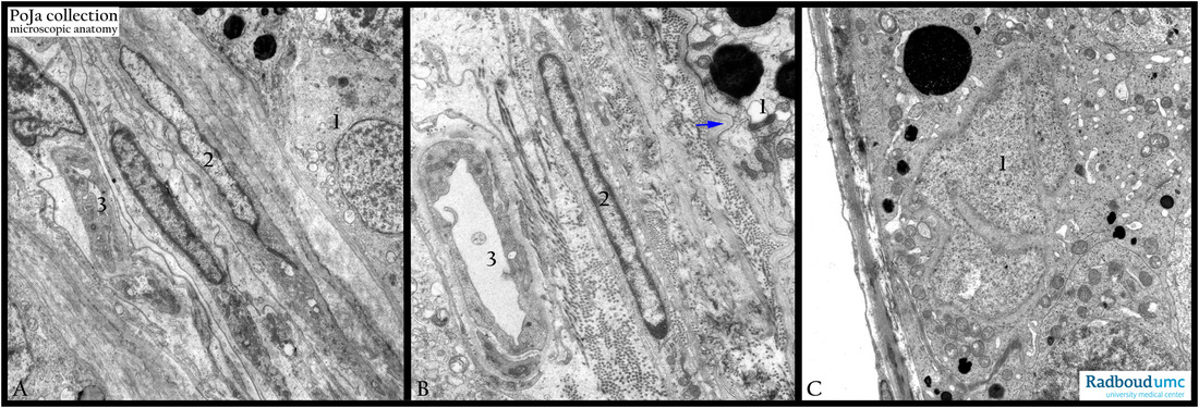
6.1 POJA-L2671+2672+2680
Title: Blood-testis barrier
Description:
(A, B) Electron microscopy, gerbil.
The myofibroblast as part of the lamina propria limitans form a barrier together with the Sertoli cells to protect the spermatogonia from possible toxic products in the bloodstream (3). (1) Sertoli cell with phagocytized electron dense material. (2) Myofibroblasts with collagen fibers in between. Note the basement membrane structure (arrows) blood-testis barrier. (3) Interstitial arteriole. Sertoli cells are activated by Follicle-stimulating hormone (FSH). They secrete inhibin and activins regulating FSH secretion. They produce androgen binding protein for stimulation of spermatogenesis and can convert testosterone into 17 beta estradiol for spermatogenesis.
Keywords/Mesh: testis, Sertoli cell, blood-testis barrier, histology, electron microscopy, POJA collection

