
12.2.4.1 POJA-La0110+L2616+
3589+2965+2618
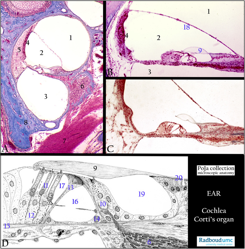
Cochlear duct and organ of Corti in the inner ear
12.2.4.1 POJA-La0110+L2616+3589+2965+2618
Title: Cochlear duct and organ of Corti in the inner ear
Description:
(A): Survey part of cochlear duct within the bony labyrinth of the cochlea, stain Azan, guinea pig.
(B): Organ of Corti, stain Azan, guinea pig.
(C): Organ of Corti, stain Haematoxylin-Weigert, human.
(D): Scheme electron microscopy organ of Corti, human.
(1) Scala vestibuli.
(2) The cochlear duct is the spiral membranous labyrinth of the cochlea (scala media) limited by the lamina basilaris (15),
stria vascularis (4) and (18) Reissner’s membrane.
(3) Scala tympani.
(4) The stria vascularis is a specialised stratified epithelium with intraepithelial capillaries. It produces endolymph that resembles intracellular fluid in ionic composition. It also regulate K+ homeostasis of the endolymph.
(5) The spiral ligament behind the stria vascularis is composed of connective tissue and functions also as the outer attachment for
the basilar membrane.
(6) Ganglion spirale. There are about 30,000 ganglia in the modiolus.
(7) Vestibulocochlear nerve.
(8) Osseous spiral membrane.
(9) Membrana tectoria. Bundles of 20 nm collagen fibrils are embedded in a tectorin-based matrix sheet with characteristic striation.
(10) Inner hair cell (IHC).
(11) Three outer hair cells (OHCs).
(12) Supportive cells (Deiters phalangeal cells).
(13) Outer pillar cell.
(14) Inner pillar cell.
(15) Lamina basilaris.
(16) Inner tunnel or tunnel of Corti with free-traversing nonmyelinated nerve fibres that run via the Nuel’s space (17) and
synapse on the basal poles of IHC and OHCs.
(19) Internal spiral tunnel.
(20) Interdental cells.
(D): The sensory cells (hair cells, 10, 11) of the organ of Corti are close associated with the supporting phalangeal cells (12).
Hair cells (HCs) are arranged characteristically in rows and possess non-motile cochlear stereocilia that are arranged in
a characteristic V-pattern (an upwards slope of short to long cilia).
Background: The tectorial membrane embeds the tips of the tall stereocilia of the hair cells (HC) and after displacement of the basilar membrane and the organ of Corti due to perilymph movement in the scala tympani depolarisation of the HCs occurs.
Ion channels at the stereocilia tips open and influx of K+ ions occurs resulting in depolarisation. In this way mechanical input by
deflection of the stereocilia leads to synaptic transmission.
The microfibrillary membrana tectoria (9) is composed of collagen types (II, V, IX, XI), glycosaminoglycans, and for 50 % of specific
tectorin and otogelin proteins. The membrane is synthesized by interdental cells (20) and extend to the external outer hair cells (OHCs).
Otogelin is a specific protein synthesized by non-sensory cells located underneath the acellular membranes such as tectorial membrane.
In maturing stages otogelin labeling is found in thick and spaced radial fibre-like structures.
Otoancorin is located at the interface between the apical surface of the inner ear sensory and non-sensory epithelia and their overlying acellular gels. In the cochlea otoancorin is detected a.o. at the attachment zones of the tectorial membrane along the top of the spiral limbus. Otoancorin ensures the attachment of the inner ear acellular gels to the apical surface of the underlying nonsensory cells.
Keywords/Mesh: inner ear, cochlear duct, organ of Corti, cochlear nerve, spiral ganglion, scala vestibuli, scala tympani, perilymph, endolymph, histology, electron microscopy, POJA collection