
7.1 POJA-L1476+L1760+L1770+L1750. Papillary serous cystadenocarcinoma of ovary (human, adult)
7.1 POJA-L1476+L1760+L1770+L1750
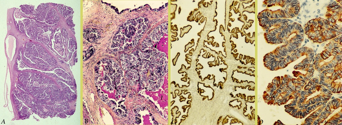
Title: Papillary serous cystadenocarcinoma of ovary (human, adult)
Description: Stain: (A) Hematoxylin-eosin; (B) Periodic acid Schiff (PAS); (C)
CK7 (OVTL12/30); (D) RCK 105 antikeratin 7 antibody immunoperoxidase staining with diaminobenzidin reaction (DAB) and hematoxylin counterstaining.
(A): Survey of well-differentiated papillary serous cystadenocarcinoma.
(B): PAS-positive secretion (1) produced by papillary tumor cells.
(C): The branched papillary projections consist of CK7-positive serous cells (OVTL12/30). Connective tissue and dilated blood vessels are negative.
(D): At higher magnification RCK 105 (CK7) reveals a heterogeneous reactivity pattern varying from distinct apical staining to moderate or minimal cytoplasm staining.
Background: Cytokeratin 7 is generally expressed by normal simple lining epithelia as well as by the epithelium of most mucous or serous glands. Generally well- differentiated malignant neoplastic cells often preserve the reactivity in contrast to anaplastic differentiated tumors. But various types of antibodies raised against the low molecular weight protein cytokeratin 7 also show different pattern of reactivities. Dependent on the cellular localization of epitopes the antibodies only recognized specific macromolecular complexes in epithelial tumor cells. Histogenetically it is assumed that a majority of epithelial tumors of the ovary is derived from the ovarian serosa. This lining is the equivalent of the embryonic coelomic epithelium in adulthood. Embryologically the coelomic epithelium give rise to the müllerian epithelium (ducts of Müller) and its derivates are the fallopian tube lining (tubal pathway), endometrial lining (endometrial pathway) and endocervical glands lining (endocervical pathway). The ovarian surface epithelium contains undifferentiated cells that possess the latent competence to differentiate along similar embryonic pathways. In this way a tumor derived from these cells will differentiate in similar way. This results in serous tumors formed by epithelial tumors that differentiate along the tubal pathway. Serous cystadenocarcinomas are essentially a malignant form of serous cystadenomas; they account for ca. 40% of all ovarian cancers and appear to be the most common malignant tumors. Mostly they are bilaterally and occurring later in life than benign and borderline tumors which usually are found between 20 and 50 years. The size of malignant serous tumors varies between10 and 15 cm but sometimes 40 cm diameters are reported. With hemorrhagic foci the tumors are mostly cystic but also partially solid. The fibrous cysts are lined by tubal-like columnar, often ciliated cells and contain blood-stained serous fluid. Solid areas of the tumor are composed of closely packed papillae that might penetrate the tumor capsule. Luxuriant papillomatous growths also extend over the surface and obliterate the ovarian structure. Destructive infiltrative stromal invasion is evident as the tumor ingrowth is diffuse or as solid cell sheets. Extreme degrees of nuclear atypia indicate progress towards undifferentiated tumor cells. Psammoma bodies (calcified concrements) though not specific might be present, too.
Keywords/Mesh: female reproductive organs, ovary, papillary serous cystadenocarcinoma, tumor keratin 7, PAS staining, female genitalia, ovarian neoplasms, histology, POJA collection