7.3 POJA-L1567+1288
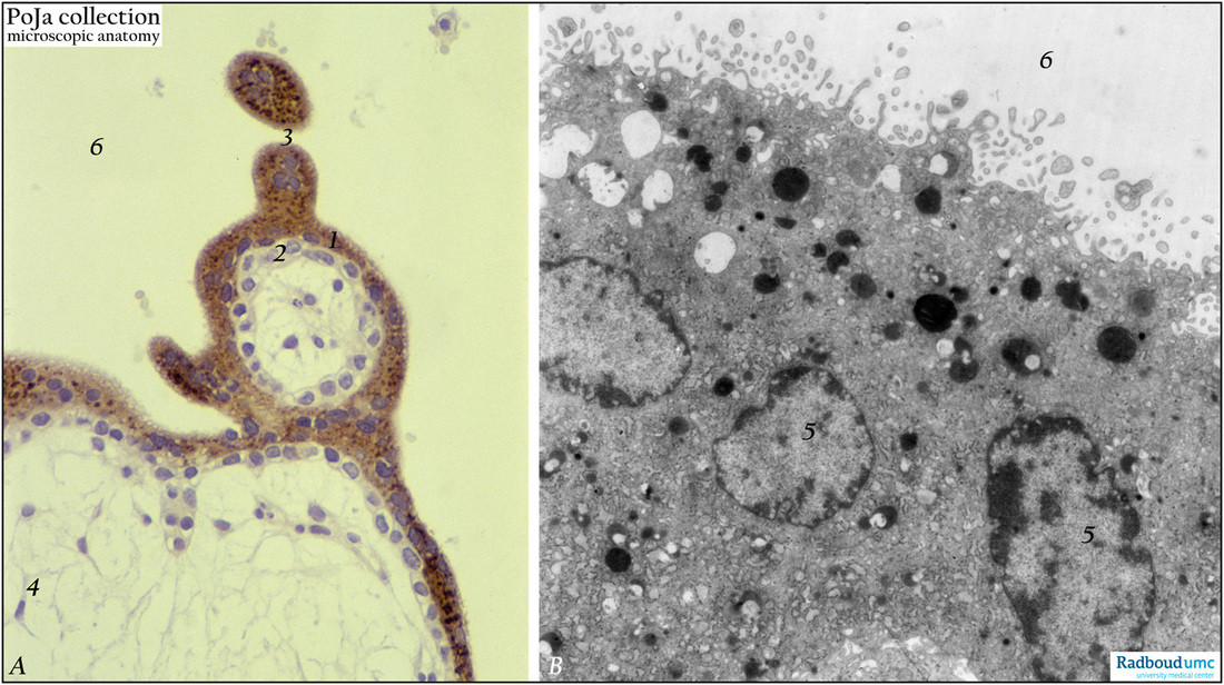
Title: Syncytiotrophoblast cells of tertiary villi (human placenta, midpregnancy)
Description: (A) Left: stain: Immunoperoxidase staining with diaminobenzidin (DAB) and human anti-chorionic gonadotrophin antibody (hCG-DAKO 231), counterstained with hematoxylin. (B) Right: electron microscopy.
(A): At the left the exclusive reactivity (dark brown) in the cytoplasma of the multinucleated syncytiotrophoblast cells (STCs, 1) is shown. The underlying cytotrophoblast cells (CTCs, 2) remain unstained and are therefore not involved in the production of hCG. The so-called syncytiotrophoblast knots and its cross-section (3) are still positive for hCG. The dark brown-stained granules reflect their lysosomal content rather than hCG. Due to the DAB-reaction an enhanced staining of the population of lysosomal granules of the STCs is obtained. (4) indicates embryonic stroma.
(B): At the right the free surface of a STC is always characterized by small protrusions being microvilli of varying length and diameter with pinocytotic invaginations. Abundant electron-dense lysosomal and light-stained endocytic structures of varying sizes are found in all STCs (nucleus, 5) and correspond to the dark brown DAB-stained granules in (A). Note that the cytoplasm is fully packed with profiles of endoplasmic reticulum indicating an intensive protein metabolism. (6) marks intervillous space in (A) and (B).
Keywords/Mesh: Syncytiotrophoblast, placenta, chorionic villi, HCG, immunohistochemistry, electron microscopy, trophoblast, histology, POJA collection.

