
11.2.1 POJA-L4433+3457+
3172+3181+3184+3185+3187.
Craniospinal ganglion (dorsal root ganglion)
11.2.1 POJA-L4433+3457+3172+3181+3184+3185+3187
Title: Craniospinal ganglion (dorsal root ganglion)
Description:
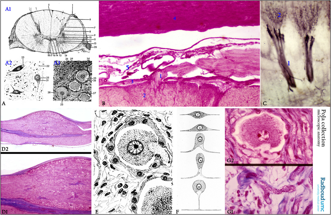
(A-A1): Scheme spinal cord and spinal medulla, human (zoom in).
Referred to Section 11.1.
(1) Radix dorsalis, dorsal root fibers.
(2) In white matter the funiculus posterior (spino-bulbar tract).
(3) Fasciculus cuneatus (column of Burdach).
(4) Fasciculus gracilis (column of Goll).
(5) Posterior median septum.
(6) Zona terminalis.
(7) Substantia gelatinosa (Rolandi).
(8) Nucleus proprius dorsalis.
(9) Formatio reticularis, reticular process and nucleus reticularis.
(10) Nucleus dorsalis (column of Clark).
(11) Lateral horn (cornu lateralis).
(12) Intermediate gray (zona intermedia).
(13) Anterior gray horn (cornu anterius).
(14) Central canal (canalis centralis).
(15) Zona intermedia centralis and lateralis.
(16) Anterior white commissure (commissura anterius alba).
(17) Anterior median fissure (fissura mediana anterius).
(18) Anterior spinal artery.
(19) Radix ventralis, anterior root fibers.
(20) Spinal ganglion.
(A-A2): Anterior gray horn (cornu ventral or anterius). (21) Motoneuron of the cornu anterius. (22) Nuclei of glia cells.
(23) Axon hillock. (25) Nucleolus.
(A-A3): Spinal ganglion. (23) Axon hillock. (24) Axons. (25) Nucleolus. (26) Endoneurium. (27) Amphicytes or mantle cells.
(28) Peripheral pseudo-unipolar ganglion cells. (29) Nucleus of the cell of Schwann.
(B): Modified silver stain, bovine. Dorsal root fibers (1). (2) White matter (posterior funiculus). (3) Pia mater.
(4) Dura mater, consisting of dense connective tissue. (5) Indicates arachnoid, being a layer of fibrocollagenous tissue covered by
flat epithelial cells (hardly visible here). This layer is not anchored to the dura mater. The space below the arachnoid or subarachnoid
space is filled with cerebrospinal fluid.
(C): Fetal mouse. Vibratome 60 µm section. Two spinal ganglia stained with immunoperoxidase and DAB and antibodies against neurofilament (NF) (1), illustrating the extremely fine arborification (2) of the nerve processes.
(D1, 2): Azan, human. Spinal ganglion (D1) together with part of the ventral root fibers (radix ventralis) (depicted in D2).
The ganglion is surrounded with a bluish stained connective tissue capsule. The large cells are the ganglion cells, embedded in a network
of myelinated nerve fibers.
(E): Scheme spinal ganglion cell with axon hillock , surrounded with small satellite cells, human.
(F): Scheme of the development of various types of polar nerve cells into a pseudo-unipolar neuron.
(G1, 2): Stain Azan, human. Spinal ganglion cell and axon hillock (G2) with part of a T-axon (G1).
Keywords/Mesh: nervous system, spinal cord, meninx, dura mater, arachnoid, pia mater, spinal ganglion cell,
dorsal root ganglion, satellite cell, nerve fiber, myelin, T-axon, neurofilament, histology, POJA collection
Title: Craniospinal ganglion (dorsal root ganglion)
Description:
(A-A1): Scheme spinal cord and spinal medulla, human (zoom in).
Referred to Section 11.1.
(1) Radix dorsalis, dorsal root fibers.
(2) In white matter the funiculus posterior (spino-bulbar tract).
(3) Fasciculus cuneatus (column of Burdach).
(4) Fasciculus gracilis (column of Goll).
(5) Posterior median septum.
(6) Zona terminalis.
(7) Substantia gelatinosa (Rolandi).
(8) Nucleus proprius dorsalis.
(9) Formatio reticularis, reticular process and nucleus reticularis.
(10) Nucleus dorsalis (column of Clark).
(11) Lateral horn (cornu lateralis).
(12) Intermediate gray (zona intermedia).
(13) Anterior gray horn (cornu anterius).
(14) Central canal (canalis centralis).
(15) Zona intermedia centralis and lateralis.
(16) Anterior white commissure (commissura anterius alba).
(17) Anterior median fissure (fissura mediana anterius).
(18) Anterior spinal artery.
(19) Radix ventralis, anterior root fibers.
(20) Spinal ganglion.
(A-A2): Anterior gray horn (cornu ventral or anterius). (21) Motoneuron of the cornu anterius. (22) Nuclei of glia cells.
(23) Axon hillock. (25) Nucleolus.
(A-A3): Spinal ganglion. (23) Axon hillock. (24) Axons. (25) Nucleolus. (26) Endoneurium. (27) Amphicytes or mantle cells.
(28) Peripheral pseudo-unipolar ganglion cells. (29) Nucleus of the cell of Schwann.
(B): Modified silver stain, bovine. Dorsal root fibers (1). (2) White matter (posterior funiculus). (3) Pia mater.
(4) Dura mater, consisting of dense connective tissue. (5) Indicates arachnoid, being a layer of fibrocollagenous tissue covered by
flat epithelial cells (hardly visible here). This layer is not anchored to the dura mater. The space below the arachnoid or subarachnoid
space is filled with cerebrospinal fluid.
(C): Fetal mouse. Vibratome 60 µm section. Two spinal ganglia stained with immunoperoxidase and DAB and antibodies against neurofilament (NF) (1), illustrating the extremely fine arborification (2) of the nerve processes.
(D1, 2): Azan, human. Spinal ganglion (D1) together with part of the ventral root fibers (radix ventralis) (depicted in D2).
The ganglion is surrounded with a bluish stained connective tissue capsule. The large cells are the ganglion cells, embedded in a network
of myelinated nerve fibers.
(E): Scheme spinal ganglion cell with axon hillock , surrounded with small satellite cells, human.
(F): Scheme of the development of various types of polar nerve cells into a pseudo-unipolar neuron.
(G1, 2): Stain Azan, human. Spinal ganglion cell and axon hillock (G2) with part of a T-axon (G1).
Keywords/Mesh: nervous system, spinal cord, meninx, dura mater, arachnoid, pia mater, spinal ganglion cell,
dorsal root ganglion, satellite cell, nerve fiber, myelin, T-axon, neurofilament, histology, POJA collection