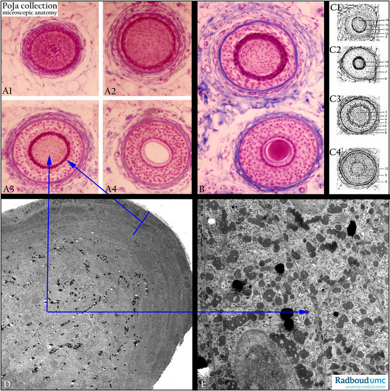
10.5 POJA-L2163+2068+2164+2175+2170
Title: Cross-sections IV through human hair at various levels
Description:
(A, B): Stain Azan. Compare photographs in (A, B) with schemes (C1-C4).
(C1 - C4): Scheme of cross-sections through hair, going from top to down.
(C1) High level: (8) Lamina membrana vitrea, glass membrane. (15) Stratum corneum.
(13) Stratum germinativum. (16) Cortex of shaft.
(14) Stratum lucidum.
(C2) Lower level: (8) Glass membrane. (16) Cortex of shaft.
(7) External root sheath. (17) Fatty degeneration of internal root sheath
(C3) Lower level: (2) Hair cortex. (6) Henle’s layer.
(3) Hair cuticle. (7) External root sheath.
(4) Sheath cuticle. (8) Glass membrane.
(5) Huxley’s layer.
(C4) Bulbus level: (8) Glass membrane.
(11) Matrix with pigmented cells.
(12) Hair papilla (dermis connective tissue).
(D, F): Electron micrographs of hair cortex with Huxley's layer. Note cross-sectioned heavy bundles of cytokeratin
(tonofilaments), they are dark electron-grey. Pigment granules are found interspersed and are -black stained.
Layer of Huxley in (D) indicated by arrow +bar is similar to (17) in (C2).
Keywords/Mesh: skin, hair, glass membrane, histology, electron microscopy, POJA collection

