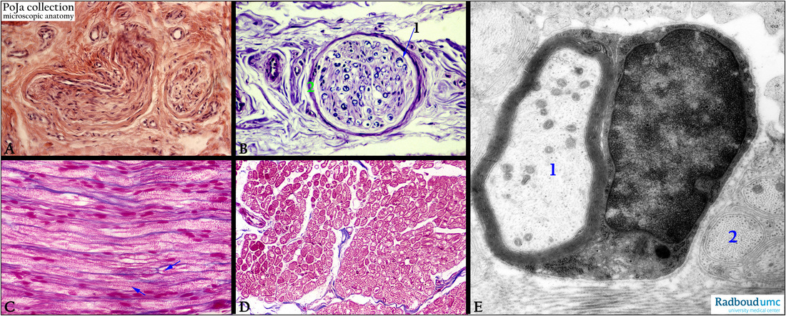
11.2 POJA-L3226+4321+3227+3228+3274
Title: Myelinated axons 2
Description:
(A): Stain hematoxylin-azophloxin, human. Myelinated nerve bundle in dermis with perineurium around it.
(B): Stain Kluver-Barrera, human. Peripheral nerve bundle with myelinated nerve fibers (1) and a perineural capsule (2), embedded in connective tissue.
(C): Stain Azan, human, longitudinal section through peripheral myelinated nerves in connective tissue. The blue arrows point to the thin axon surrounded by a thick layer of granular looking myelin sheath. This sheath is composed of concentrically wrapped Schwann cell membranes. Note also the nodes of Ranvier (near the arrow).
(D): Stain Azan, human. Cross-section equivalent to (C).
(E): Electron micrograph, adrenal gland, rat. A single Schwann cell with a thick electron-dense myelin wrap around one axon (1).
The axon displays distinctly neurotubuli and neurofilaments. Compare also to cross-sections of nonmyelinated axons (2) at the right side.
A thin basal lamina or external lamina always surrounds each cell of Schwann.
Keywords/Mesh: nervous tissue, axon, peripheral nerve fiber, myelinated nerve fiber, Schwann cell,
node of Ranvier, perineurium, histology, electron microscopy, POJA collection

