5.4.3 POJA-L5002+2432+La0354+La0056+4257+2461+4262+2462
Title: Proximal and distal convoluted tubules (X) in the kidney
Description:
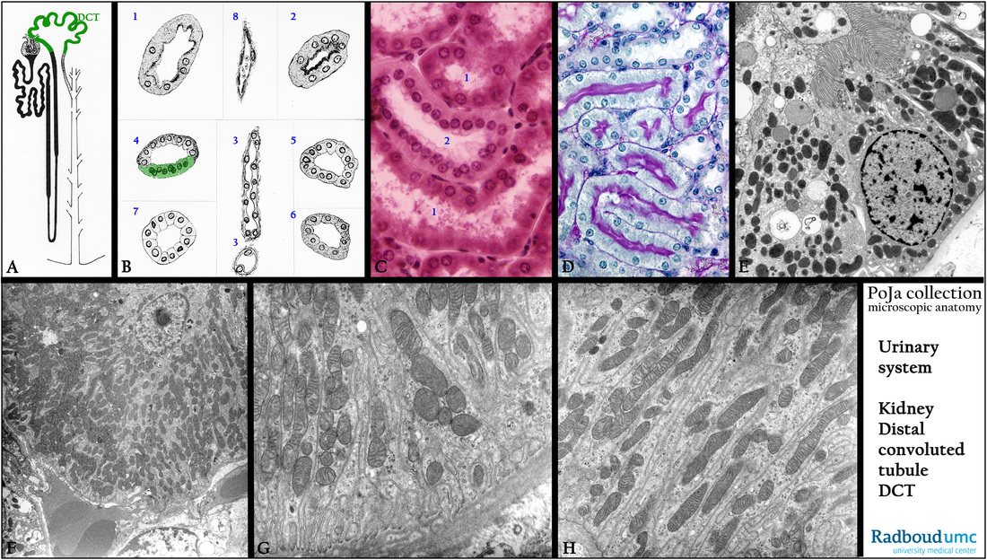
(A): Scheme, nephron, human. The green painted loop represents the distal convoluted tubule (DCT).
(B): Scheme of cross-sections through various types of renal tubules, human:
(1) Pars recta I. (5) Pars contorta II.
(2) Pars contorta I. (6) Pars recta II.
(3) Henle’s loop (2x). (7) Collecting tube.
(4) Macula densa (green). (8) Capillary.
(C): Kidney cortex, stain Weigert hematoxylin-azophloxine, rat. (1) Proximal straight tubule (PST). (2) Cortical thick ascending limb (cTAL).
(D): Kidney cortex, stain PAS-hematoxylin staining, rat. The proximal tubules (PST) are studded with PAS-positive microvilli on the cell surface. Note that also the basal lamina is positive. The distal tubules are negative.
(E): Electron microscopy, human. A PCT (proximal convoluted tubule) , illustrating a cell type with numerous dense-stained mitochondria and grey-stained round lysosomes. Apically interwoven long microvilli (brush border) and many small endocytotic structures.
(F): Electron microscopy, human. Distal tubules (DCT) close to the glomerulus, illustrating a cell type with numerous mitochondria
and hardly lysosomes.
(G, H): Electron microscopy, human. Details of the basal labyrinth of DCTs showing characteristic arrangement of mostly long, slender mitochondria between the deep basal infoldings of the cell membrane.
Keywords/Mesh: urinary system, kidney, distal convoluted tubule, proximal convoluted tubule, histology, electron microscopy, POJA collection

