2.2 POJA –L1006
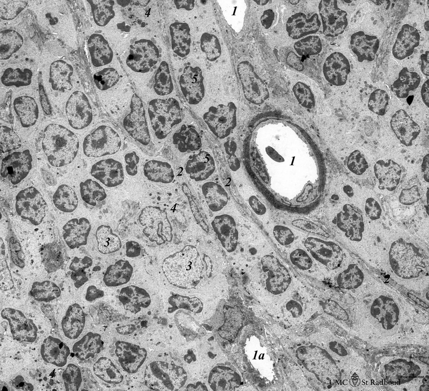
Title: Survey electron microscopy of part of a lymphatic nodule (rat)
Description: Electron microscopy.
(1) Shows an arteriolar branch of a central artery, and cross-sections of capillary branches (1a). Within the periarteriolar lymphatic sheath (PALS) mostly T-lymphocytes (5) are present between concentric arranged reticular cells (2). Cells with larger lighter stained nuclei represent dendritic cells (APC) (3). Few macrophages are also present (4).
Keywords/Mesh: lymphatic tissue, spleen, central artery, histology, electron microscopy, POJA collection

