2.1 POJA-L937
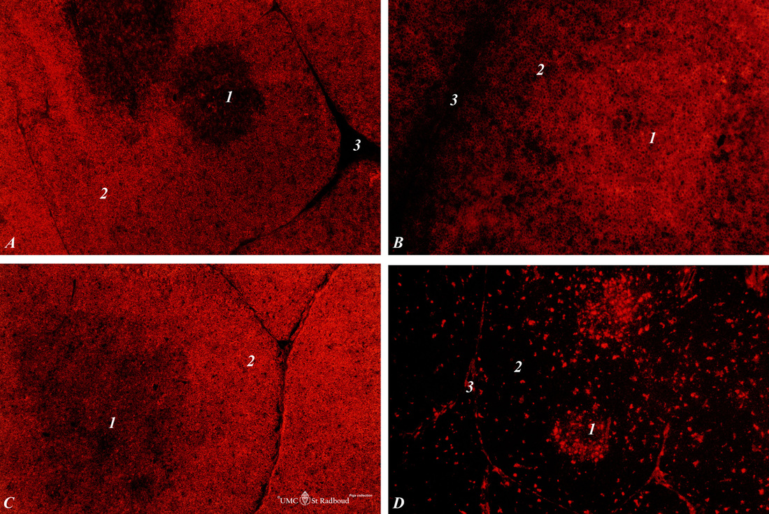
Title: Immunohistochemistry of subsets of T cells and macrophages marked by CD8-, OX19-, ER1- and ED1 markers in normal thymus (rat)
Description: Stain: Alexa 594 red immunofluorescence.
(1): Medulla;
(2): Cortex;
(3): Septa
(A): Strong CD8 staining of the thymic cortical lymphocytes (single CD8+ and double positive CD4+CD8+ thymocytes).
(B): OX19 antibody staining for almost all T cells, equivalent to T1 human marker and Ly-1 mouse marker. Note that the (mature) medullary T cell show stronger OX-19 expression than the (immature) cortical thymocytes.
(C): ER1 antibody stains all T cells being equivalent to CD43. Both cortex and medulla are well stained.
(D): ED1 antibody stains resident tissue macrophages which are present more in the well capillarised medulla than in the cortical area.
Keywords/Mesh: lymphatic organs, thymus, immunohistochemistry, CD8, OX19, ER1, ED1, rat, toxicology, histology, POJA collection

