
5.4.1 POJA-L2467+2378+
2468+2537+2542.
The Bowman capsule of the glomerulus (IX) in the kidney
5.4.1 POJA-L2467+2378+2468+2537+2542
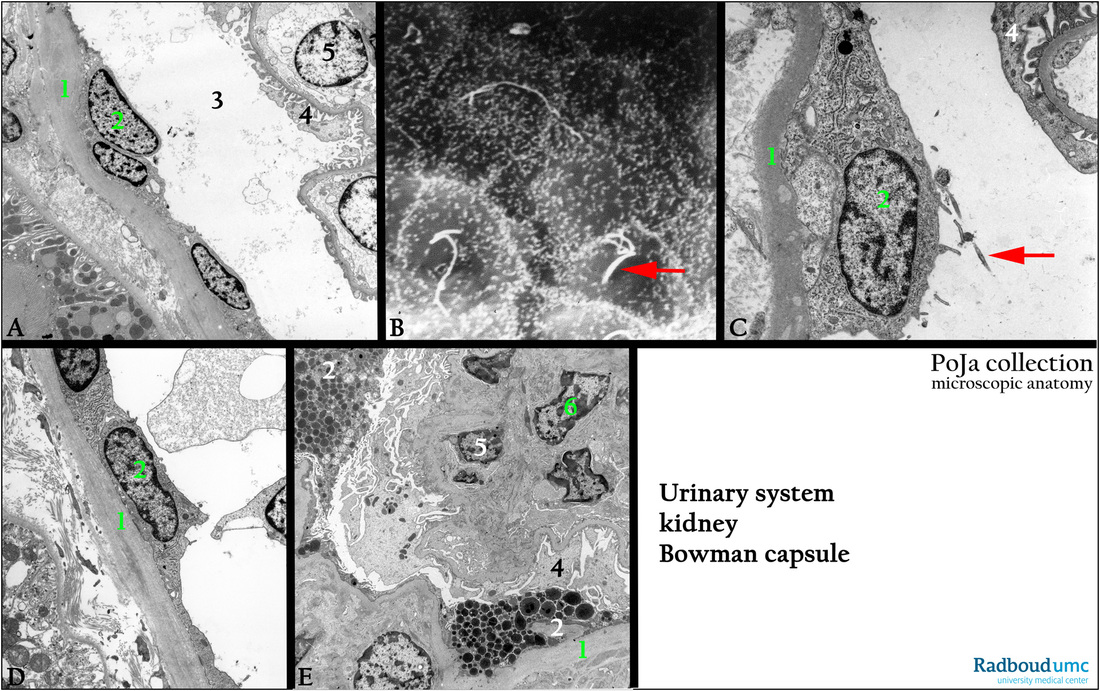
Title: The Bowman capsule of the glomerulus (IX) in the kidney
Description:
(A, C, D): Glomerulus, electron microscopy, human.
(1)Thick basal lamina of Bowman’s capsule.
(2) Parietal cell with (kino)cilium (red arrow) and short microvilli. These cells can easily proliferate, detach and even phagocytize.
(3) Bowman’s space.
(4) Pedicles of podocyte surrounding the blood vessel.
(5) Capillary, marked by the endothelial cell.
(B): Bowman capsule, scanning electron microscopy, rat. Parietal cells (2) with short microvilli and one single central cilium
(white long thread, red arrow).
(E): Glomerulus, electron microscopy, human. (2) Phagocytizing parietal cells. (6) Mesangium cell.
Keywords/Mesh: urinary system, kidney, glomerulus, parietal cell, Bowman’s capsule, histology, electron microsocopy, POJA collection