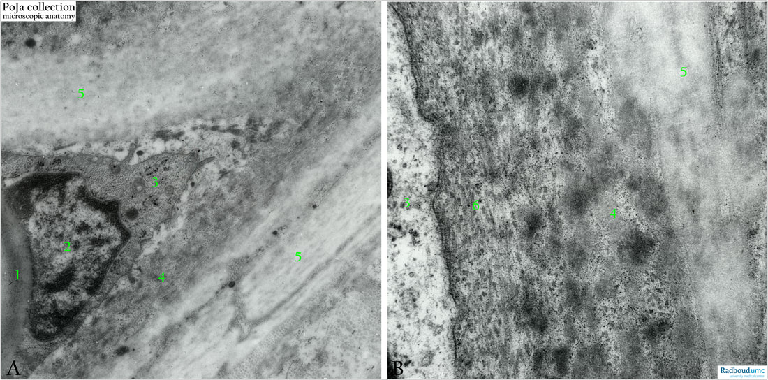
15.2 POJA-L7032+7035 Electron micrographs of elastic cartilage
15.2 POJA-L7032+7035 Electron micrographs of elastic cartilage
Title: Electron micrographs of elastic cartilage
Description:
Ultrastructural low magnification (A) and detail (B) of the ultrastructure of elastic cartilage in the auricle (gerbil)
(1): Lipid (partly visible) in the chondrocyte.
(2): Nucleus.
(3): Cytoplasm.
(4): In the vicinity of the chondrocyte cytoplasm (3) long thin collagen fibrils course between numerous deposits of newly formed elastin.
In (B) the enlarged denser fluffy spots of new elastin are well visible. The immature elastin also referred as oxytalan fibril is presented as dense tangled deposit of elastin protein surrounded by short microfibrils e.g. fibrillin, (See also: Respiratory System 8.5 POJA-L467+451+465 Detail elastin in alveolar tip).
(5): Amorphous elastin; lower bottom (A-5) mature elastin between thin branching processes of chondrocyte.
(6): Thin collagen fibrils.
Background:
Ultrastructurally a light microscopic elastic fibre is composed of amorphous elastin surrounded by and associated with microfibrils (diameter 10-11 nm). It appear that during development firstly the microfibrils are produced and laid down. Subsequently the elastin is deposited within a microfibrillar network that serves as a scaffold for the lay down of elastin. It is assumed that oxytalan fibrils or fibres form a developmental stage of the mature elastin.
Keywords/Mesh: locomotor system, cartilage, elastic, matrix, chondrocyte, oxytalan fibril, elastic fibril, elastin, collagen fibril, electron microscopy, POJA collection

