4.1.1 POJA-L4034+4035
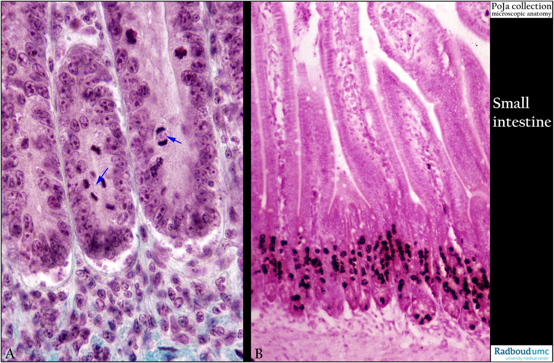
Title: Dividing cells in crypts of small intestine (mouse, dog)
Description: Stain: (A) Trichrome-Goldner, dog. (B) Tritium thymidine labeling-and counterstained with eosin, mouse.
(A): Most mitoses are found in the lower zone (arrows, ↘↘) of the crypts of the small intestine. After mitosis cells move upwards and differentiate to resorptive cells and mucus producing cells as well as enteroendocrine cells.
(B): Tritiated thymidine is incorporated in dividing, mitotic cells, the label is visualized by autoradiography, and the slide confirms that dividing cells and DNA-synthesizing cells are preferentially found in the lower zone of the intestinal crypts.
Keywords/Mesh: small intestine, crypts, mitosis, cell proliferation, 3H-thymidine labeling, autoradiography, histology, POJA collection

