
13.1 POJA-L4534+4536+4535+4539+4538 Purkinje fibres in the heart
13.1 POJA-L4534+4536+4535+4539+4538
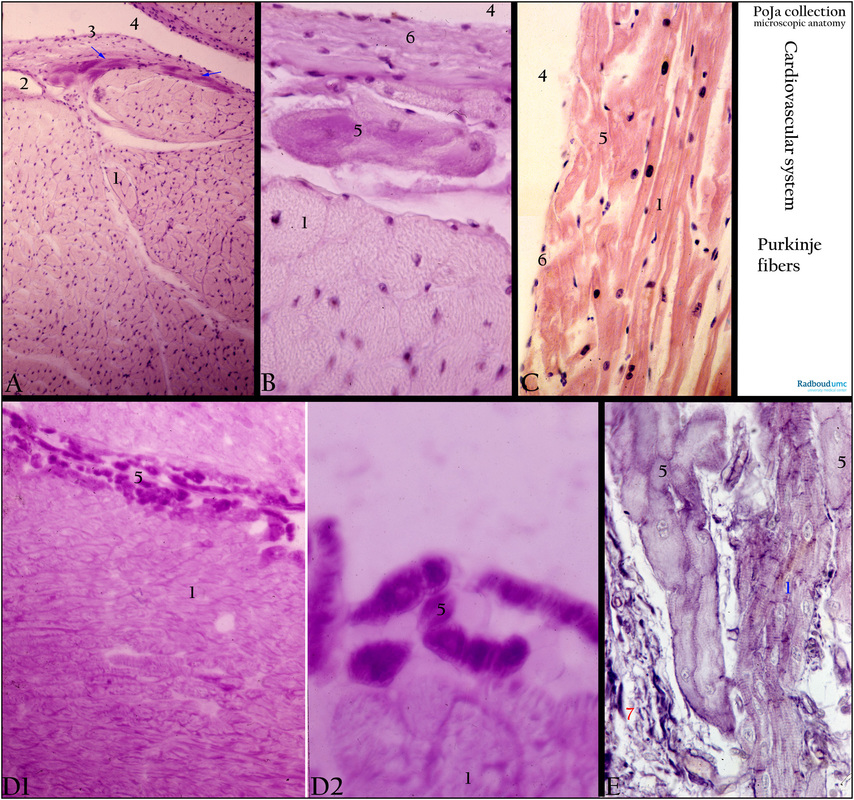
Title: Purkinje fibres in the heart
Description:
(A): Heart, ventricle, conducting system, PAS, human. Specialised modified heart cells (so-called nodal cardiocytes) in branches of the conducting system also referred as Purkinje fibres. These elongated cylindrical cells are PAS positive (presence of glycogen) and form strands (arrows) below the subendocardial tissue. (1) Normal or common myocardiocytes (so-called contractile cardiocytes). (2) Blood vessel. (3) Endothelial cells of endocardium. (4) Lumen ventricle.
(B): Heart, ventricle, conducting system, PAS, human. Specialised modified cardiomyocytes (nodal cardiocytes) in branches of the conducting system also referred as Purkinje fibres (5). These elongated cylindrical cells are PAS positive and form strands below the subendocardial tissue (6). (1) Common contractile cardiocytes, (4) lumen ventricle.
(C): Heart, ventricle, conducting system, HE, human. Specialised modified heart cells (nodal cardiocytes) in branches of the conducting system also referred as Purkinje fibres (5). These elongated cylindrical cells form strands below the endocardial lining (6). (1) Common cardiocytes (so-called contractile cardiocytes). (4) Lumen ventricle and endothelial cells of endocardium (6).
(D1/2): Survey and detail of heart, ventricle, conducting system, PAS, rat. In the wall of the ventricle a network of terminal branches (5, Purkinje fibres) of the conducting system is distinctly PAS positive. These modified cardiomyocytes (nodal cardiocytes) arranged as cord-like clusters (5) run between the common contractile cardiocytes and contain a large amount of glycogen. Sarcolemmae (= cell membranes with external lamina) of all cardiomyocytes also stain positively. In contrast to human Purkinje fibres (5) in rat the sizes of these specialised cells do not exceed those of common contractile cardiocytes (1).
(E): Heart, ventricle, conducting system, Haematoxylin (Weigert), human. Terminal ramifications of the branching conducting system within the walls of the ventricles show elongated cylindrical cells referred as Purkinje fibres (5). These nodal cardiocytes contain central nuclei, myofibrils and light-stained areas due to the presence of glycogen. There are no intercalated discs in contrast to the adjacent common fusiform cardiomyocytes (1). Connective tissue (7) with small blood vessel.
Background: The conducting system of the heart (mammals) is composed of: 1. Sinoatrial (SA) node of Keith and Flack, 2. Atrioventricular (AV) node of Aschoff-Tawara, 3. Atrioventricular or common bundle of His, 4. Right and left branches on either side of the interventricular septum, 5. Terminal endings of the branches to the papillary muscles and the ventricle walls. The conducting cells (nodal cardiocytes) form strands supplied by many sympathetic and parasympathetic nerve fibres and capillaries. These specialised fusiform cells are modified smaller cardiocytes. They contain myofibrils and a large amount of glycogen but lack the transverse tubules of the T-system and there are no intercalated discs. The strand cells are interconnected by junctional contacts of the same type as in normal myocardiocytes. In the distal projections of the conducting system i.e. the interventricular branches the cells become larger (up to 50 micrometers width and 100 micrometers long) and are called Purkinje fibres.
The sinoatrial node (SAN) cells are also described as “pacemaker” or “nodal-like” cells considered as therapeutic potential stem cells. (doi: 10.1124/pr.114.009597 Pharmacological Reviews April 2015 vol. 67 no. 2 368-388. http://pharmrev.aspetjournals.org/content/67/2/368.long#title1).
Keywords/Mesh: cardiovascular system, heart, conducting system, Purkinje fibre, bundle of His, ventricle, myocardiocyte, cardiocyte, glycogen, sino-atrial cell, nodal cardiocyte, histology, POJA collection