
12.2.4.2 POJA-L2637+4461B+
4462
Ductus endolymphaticus of the inner ear
12.2.4.2 POJA-L2637+4461B+4462
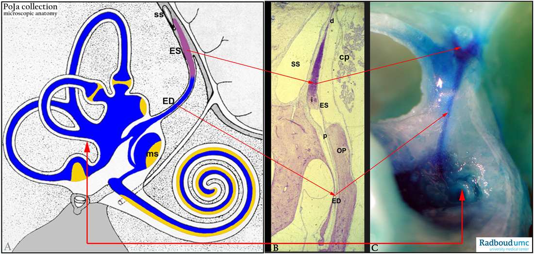
Title: Ductus endolymphaticus of the inner ear
Description:
(A): Scheme inner ear, human (by courtesy of W. Kuipers, PhD, former Head Biological Laboratory, Department of Otorhinolaryngology,
Radboud university medical center, Nijmegen, The Netherlands).
(B, C): Endolymphatic duct, stain Haematoxylin-eosin, rat.
The endolymph produced by the stria vascularis is drained from the utricle and saccule by the endolymphatic duct (ED)
to the endolymphatic sac (ES). The endolymph is resorbed through the wall of the sac into the vessels of the dura mater.
The endolymphatic sac is divided in a thin proximal part continuous with the endolymphatic duct, a wider intermediate part and a thin distal part. The latter lies close to (SS) in (B), the so-called sinus sigmoideus (or sigmoid sinus, it is a dural venous sinus that receives blood from the transverse sinuses as well as from a.o. the cerebral, cerebellar veins).
(SS) = sigmoid sinus.
(ES) = endolymphatic sac.
(d) = distal part of the endolymphatic sac.
(i) = intermediary part of the endolymphatic sac.
(p) = proximal part of endolymphatic sac.
(ED) = endolymphatic duct.
(OP) = ostia operculum.
(Cp) = choroid plexus brain.
(ms) = macula sacculi.
Vital staining with trypan blue through the sacculus (in C). The endolymph is transported to and removed in the endolymphatic sac.
The distal part of the (ES) possesses the capacity for expansion and thus may play a role in the cushioning of fluctuations in endolymph pressure.
(By courtesy of T. Peters, PhD and W. Kuipers, PhD, Department of Otorhinolaryngology, University Medical Centre of the Radboud University, Nijmegen, The Netherlands).
Background:
The lining cells of the endolymphatic sac (ES) are the light and dark cells involved in transport activities.
The light cells also show secretory activities as indicated by the presence of cytokeratin 7 (glandular, secreting cells), high concentrations of oxidative enzymes cytochrome oxidase and succinodehydrogenase (rich in mitochondria).
The distal extraosseus part of the (ES) show strong co-expression of vimentin and different types of cytokeratins conferring to
viscoelastic properties of this part. It is also indicated that this distal part of the (ES) possesses the capacity for expansion and its role is
the cushioning of fluctuations from endolymph pressure.
Meniere's syndrome (idiopathic endolymphatic hydrops) is a disease of the internal ear affecting hearing and equilibrium.
Dizziness, tinnitus and a low-frequency hearing loss occur. It is caused by an increase in endolymphatic pressure with swelling of the membranous labyrinth due to e.g. blockage of the endolymphatic duct. It is clear that the endolymphatic sac forms an essential link in the control of endolymph composition through its resorption and secretory capacities.
Keywords/Mesh: inner ear, endolymphatic sac, endolymphatic duct, vestibulum, semicircular canal, histology, POJA collection