12.2.4.2 POJA-L3617+3449+3448+3613+3618+3630
Title: Planum semilunatum of the saccule in the inner ear
Description:
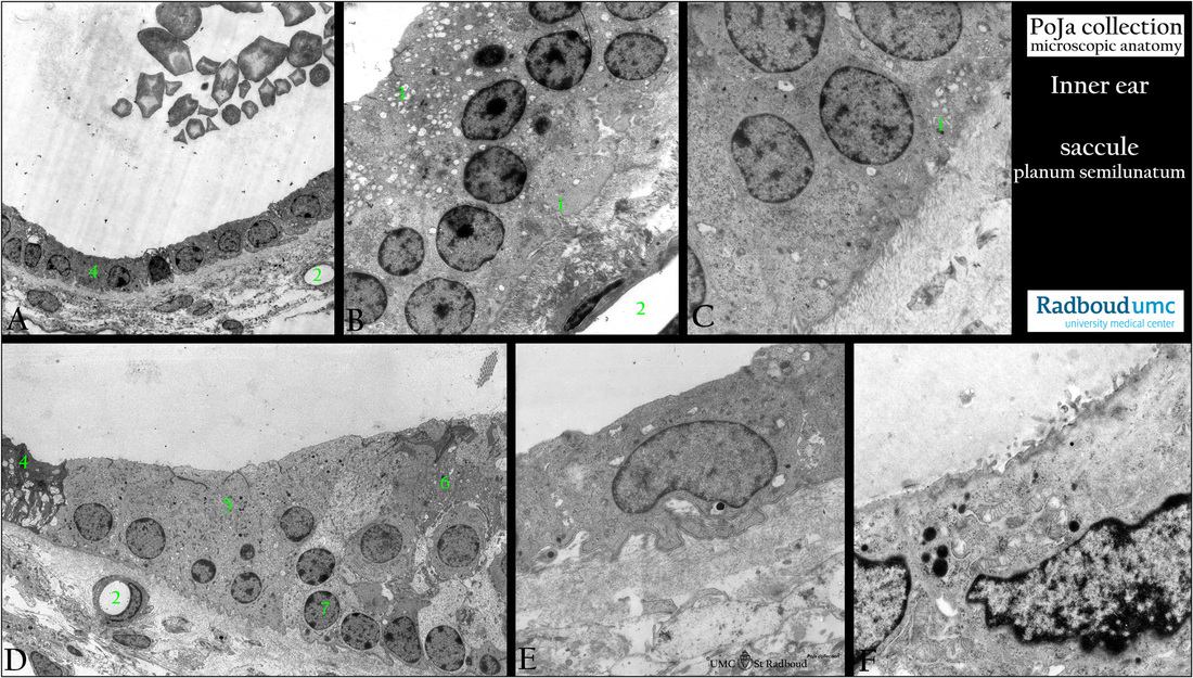
(A - F): Electron micrographs of saccule and part of the planum semilunatum, rat.
At the top in a macula the sensory epithelium of hair cells and supporting cells and downwards toward the base of the crista and
macula the transition into the planum semilunatum. Around the base of the cristae and maculae the planum semilunatum is an area
that is composed of a single lining of non-sensitive epithelium. The cells are non-ciliated cuboidal to flattened and continuous with
regular lining of the membranous semicircular canal.
Background:
The cells in the planum semilunatum are thought to be involved in secretory functions, additionally protein-secreting structures are
found located in the apical region of the cells. A subpopulation of cells also appear to contain carboanhydrase that is involved in the secretion of endolymph K+ as well as in regulating fluid balance. Vessels beneath the area of the planum semilunatum are found to
form a specific loop-shaped route most likely involved in secretion and reabsorption of endolymph.
(A): Low magnification also shows otoconia in the lumen of the saccule.
(B-F): Lining epithelial cells of the peripheral part of the macula exhibit different cellular features.
The morphological characteristics indicate a secretory and transport function of these cells.
(B) Cytoplasmic vacuoles (3) in these lining cells, presence of basal cell membrane indentations (1). Note subepithelial capillary (2).
(C) Basally small indentations (1) in peripheral supporting cells of the macula.
(D) Peripheral located dark cells with vacuoles and basal and lateral deep foldings (4), similar as in (A, 4).
(5) Electron light cells being a transitional epithelium with small basal invaginations and lateral interdigitations.
(6) Last hair cells and supporting cells (7) located below the hair cells. The cells in 4+5 are called together planum semilunatum.
(2) Note always presence of subepithelial capillaries.
(E) Peripheral lining flattened cell with broad leaflet-like basal cell membrane folding as part of the planum semilunatum.
(F) Peripheral supporting cell with electron-dense secretory granules in the apical region. It is assumed that the cells are involved in secretory functions and on their account are also the regulation of the endolymph volume and transport of H2O.
They secrete sulfated proteoglycans and distinct Na-K-ATPase is detected in the cells. The electron-dense cells play a role in the
regulation of the ionic composition of the endolymph as carboanhydrase activity is also described in their basal infoldings.
Keywords/Mesh: inner ear, vestibular organ, saccule, planum semilunatum, histology, electron microscopy, POJA collection

