
12.1.4 POJA-L4425+4426+
3568+4427
Optic nerve in the eye
12.1.4 POJA-L4425+4426+3568+4427
Title: Optic nerve in the eye
Description:
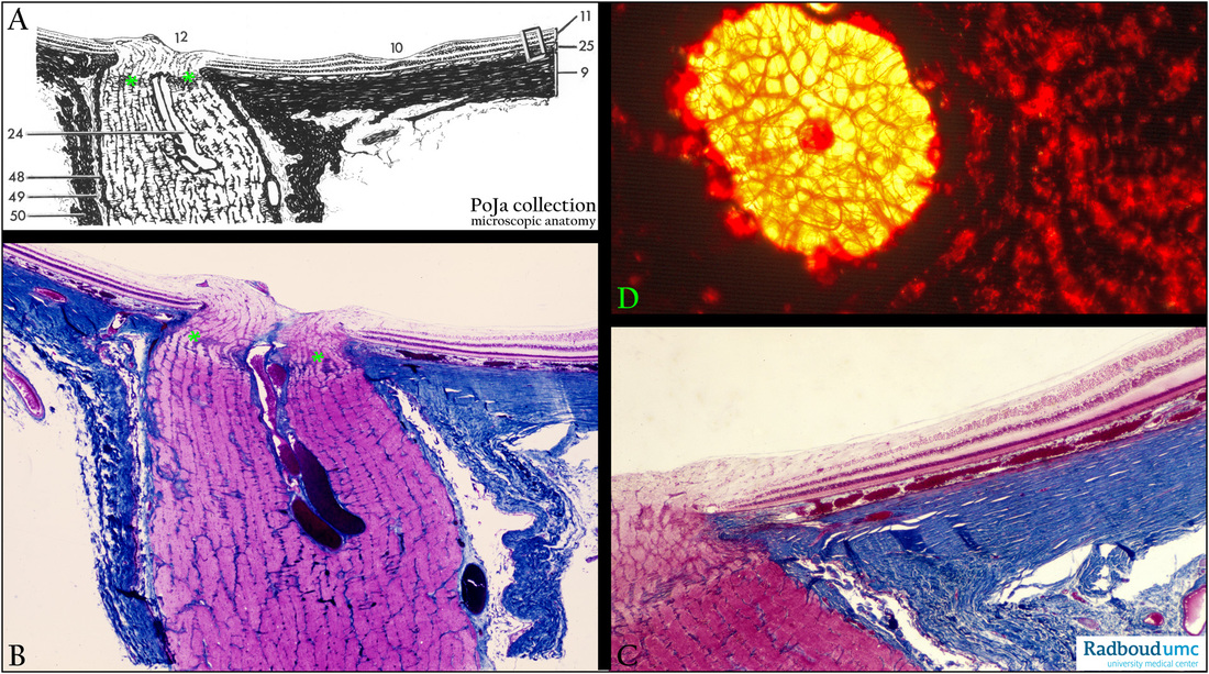
(A): Scheme optic nerve and macula lutea, human.
(9) Sclera.
(10) Macula lutea.
(11) Pars optica retinae.
(12) Papilla nervus optici (optic disc).
(24) Arteria and vena centralis retinae.
(25) Choroidea.
(48) Pia mater.
(49) Arachnoid.
(50) Dura mater.
(B, C): Optic nerve (compare with A), stain Azan, human.
Meridional section of optic nerve and optic papilla (optic disc): Identical layers and areas as in scheme (A).
The optic disc (=blind spot of the retina) is not suitable for vision as the photoreceptor cells terminate at its edges.
The disc includes the optic papilla (a protrusion formed by axons entering the optic nerve; the central depression is the optic cup) and
the lamina cribrosa (green **** in A, B) of the sclera through which the axons pass.
The intraocular part of the optic nerve is very short and formed by nonmyelinated axons of the retinal ganglion cells i.e. optic nerve fibres. The orbital part of the optic nerve is ca. 3 mm long behind the lamina cribrosa and the nerve fibres are myelinated by oligodendrocytes.
The central retinal arteria and vein are found in the middle of the optic nerve and pass through the optic disc.
The optic nerve is completely enclosed by the dura mater, arachnoid and pia mater and connects the eyeball via the chiasma opticum
with the cerebrum.
(D): Uvea in situ, Sirius red perfusion, human. The uvea or the pigmented vascularised middle tunic is divided into the choroid, ciliary
body and iris. In situ perfusion of the uvea shows pattern of reddish-stained long choroidal venous branches close to the bright yellow-
red orifice of the formerly located optic disc.
Keywords/Mesh: eye, optic nerve, retina, optic papilla, optic disc, lamina cribrosa, histology, POJA collection
Title: Optic nerve in the eye
Description:
(A): Scheme optic nerve and macula lutea, human.
(9) Sclera.
(10) Macula lutea.
(11) Pars optica retinae.
(12) Papilla nervus optici (optic disc).
(24) Arteria and vena centralis retinae.
(25) Choroidea.
(48) Pia mater.
(49) Arachnoid.
(50) Dura mater.
(B, C): Optic nerve (compare with A), stain Azan, human.
Meridional section of optic nerve and optic papilla (optic disc): Identical layers and areas as in scheme (A).
The optic disc (=blind spot of the retina) is not suitable for vision as the photoreceptor cells terminate at its edges.
The disc includes the optic papilla (a protrusion formed by axons entering the optic nerve; the central depression is the optic cup) and
the lamina cribrosa (green **** in A, B) of the sclera through which the axons pass.
The intraocular part of the optic nerve is very short and formed by nonmyelinated axons of the retinal ganglion cells i.e. optic nerve fibres. The orbital part of the optic nerve is ca. 3 mm long behind the lamina cribrosa and the nerve fibres are myelinated by oligodendrocytes.
The central retinal arteria and vein are found in the middle of the optic nerve and pass through the optic disc.
The optic nerve is completely enclosed by the dura mater, arachnoid and pia mater and connects the eyeball via the chiasma opticum
with the cerebrum.
(D): Uvea in situ, Sirius red perfusion, human. The uvea or the pigmented vascularised middle tunic is divided into the choroid, ciliary
body and iris. In situ perfusion of the uvea shows pattern of reddish-stained long choroidal venous branches close to the bright yellow-
red orifice of the formerly located optic disc.
Keywords/Mesh: eye, optic nerve, retina, optic papilla, optic disc, lamina cribrosa, histology, POJA collection