12.1.2 POJA-L2536+La0006+2958+3561
Title: Cornea of the eye
Description:
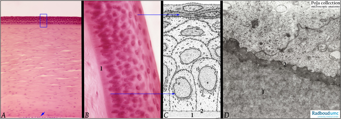
(A, B): Cornea, stain Haematoxylin-eosin, human. Survey and detail. Below (A, arrow) at the inner side of the cornea, the Descemet’s membrane limits the corneal stroma by a single layered corneal endothelium.
(C, D): Corneal epithelium, electron microscopy scheme and electron micrograph, human. Ultrastructure basal part of corneal basal cells and Bowman’s layer (D). The cornea is covered with a five to six layers of non-keratinizing squamous epithelium resting on a basal lamina firmly attached to the specialised outer condensed layer of the corneal stroma the so-called Bowman’s layer (8-15 µm), detailed in (B) and (C).
(B, C, D, 1) Bowman’s layer. The basal lamina (C, D, 2) and hemidesmosomes are present in (D).
Background: The cornea is transparent, viscoelastic, avascular and resistant to deformations. These properties are obtained a.o. by a characteristic arrangement and aligning of collagen fibres, glycosaminoglycans, affinity to water. The corneal stroma (500 µm) consists of a thick lamellar layer of parallel arranged collagen fibres with in between flattened specialised fibroblasts (or keratocytes).
Keywords/Mesh: eye, cornea, Bowman’s layer, histology, electron microscopy, POJA collection

