
9.3 POJA-L2812+2813+3497
+3496.
Calcitonin cells (C-cells) in thyroid gland (X)
9.3 POJA-L2812+2813+3497+3496
Title: Calcitonin cells (C-cells) in thyroid gland (X)
Description:
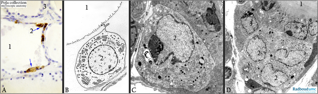
Thyroid gland. (A): Immunoperoxidase-DAB staining with anticalcitonin antibodies, human. The follicle (1) contains some C-cells stained specifically with anti-calcitonin antibodies (2). (3) Represents the regular follicular epithelial cells producing thyroglobulin.
(B): Electron microscopy scheme of a C-cell, human. The schematic structure of the calcitonin cells shows that the cell is sealed off
from the follicle space, contains numerous mitochondria and many small electron-dense granules.
(C): Electron micrograph of a C-cell, gerbil. (2) is the C-cell.
(D): Electron micrograph of C-cells and follicular cell, gerbil. A cluster of three C-cells in (2). C-cells might contain lipofuscin granules
and small lysosomes.
Keywords/Mesh: thyroid gland, follicle, follicular cell, parafollicular cell, calcitonin cell, C-cell, calcitonin, thyroglobulin, histology, electron microscopy, POJA collection
Title: Calcitonin cells (C-cells) in thyroid gland (X)
Description:
Thyroid gland. (A): Immunoperoxidase-DAB staining with anticalcitonin antibodies, human. The follicle (1) contains some C-cells stained specifically with anti-calcitonin antibodies (2). (3) Represents the regular follicular epithelial cells producing thyroglobulin.
(B): Electron microscopy scheme of a C-cell, human. The schematic structure of the calcitonin cells shows that the cell is sealed off
from the follicle space, contains numerous mitochondria and many small electron-dense granules.
(C): Electron micrograph of a C-cell, gerbil. (2) is the C-cell.
(D): Electron micrograph of C-cells and follicular cell, gerbil. A cluster of three C-cells in (2). C-cells might contain lipofuscin granules
and small lysosomes.
Keywords/Mesh: thyroid gland, follicle, follicular cell, parafollicular cell, calcitonin cell, C-cell, calcitonin, thyroglobulin, histology, electron microscopy, POJA collection