2.1 POJA-916
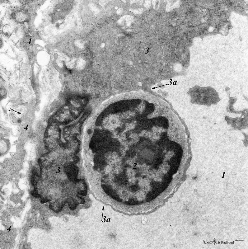
Title: Thymus cortex (rat, young adult)
Description: Electron microscopy.
Type I epithelioreticular cells (4) separate connective tissue compartment (capsule, trabeculae, blood vessels) from the thymic parenchyma.
At the top the capsule is bordered by a basal lamina (4a) of two projections (4) of type I epithelioreticular cells. Close to them, part of an endothelial cell (3) of a capillary (1: lumen). A thymocyte (2) in diapedesis through the endothelial cytoplasm is enclosed by endothelial cell processes (3a).
Keywords/Mesh: lymphatic tissue, thymus, epithelioreticular cell I, histology, electron microscopy, POJA collection

