
13.1 POJA-L4627+4629+4628+4630
Immunohistochemistry of vascular smooth muscle cells, i.e. actin, vimentin, desmin
13.1 POJA-L4627+4629+4628+4630
Title: Immunohistochemistry of vascular smooth muscle cells
Description:
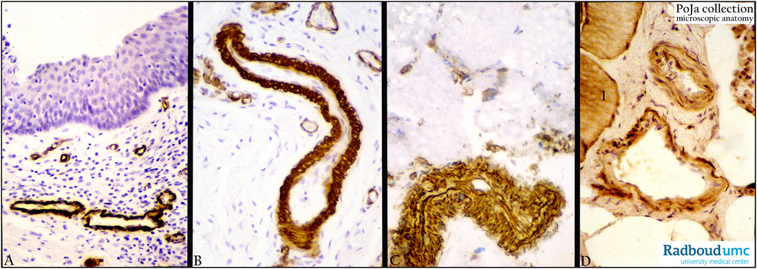
(A, B): Survey and detail of immunoperoxidase staining with DAB for α-SM-actin in the intermediate zone of the anal region (pig). The squamous surface cells are negative. Actin is found in the smooth muscle cell wall of the small blood vessels, including the pericytes. The thinner venules with wide lumen show weaker staining (in B).
(C): Immunoperoxidase staining with DAB for vimentin (anal region, pig). Vimentin is present in the smooth muscle cells and the endothelium of the artery and small capillaries.
(D): Immunoperoxidase staining for desmin (muscle biopsy, human) in small artery and vein (with larger lumen). Note that desmin is also found in the skeletal muscle fibres (1). The large fat cells are negative.
Keywords/Mesh: cardiovascular system, vascularisation, blood vessel, arteriole , muscular artery, smooth muscle, actin, vimentin, desmin, histology, POJA collection
Title: Immunohistochemistry of vascular smooth muscle cells
Description:
(A, B): Survey and detail of immunoperoxidase staining with DAB for α-SM-actin in the intermediate zone of the anal region (pig). The squamous surface cells are negative. Actin is found in the smooth muscle cell wall of the small blood vessels, including the pericytes. The thinner venules with wide lumen show weaker staining (in B).
(C): Immunoperoxidase staining with DAB for vimentin (anal region, pig). Vimentin is present in the smooth muscle cells and the endothelium of the artery and small capillaries.
(D): Immunoperoxidase staining for desmin (muscle biopsy, human) in small artery and vein (with larger lumen). Note that desmin is also found in the skeletal muscle fibres (1). The large fat cells are negative.
Keywords/Mesh: cardiovascular system, vascularisation, blood vessel, arteriole , muscular artery, smooth muscle, actin, vimentin, desmin, histology, POJA collection