14.1 POJA-L6058+6259+6145B Ultrastructure of triad system in skeletal muscle fibres
14.1 POJA-L6058+6259+6145B Ultrastructure of triad system in skeletal muscle fibres
Title: Ultrastructure of triad system in skeletal muscle fibres
Description:
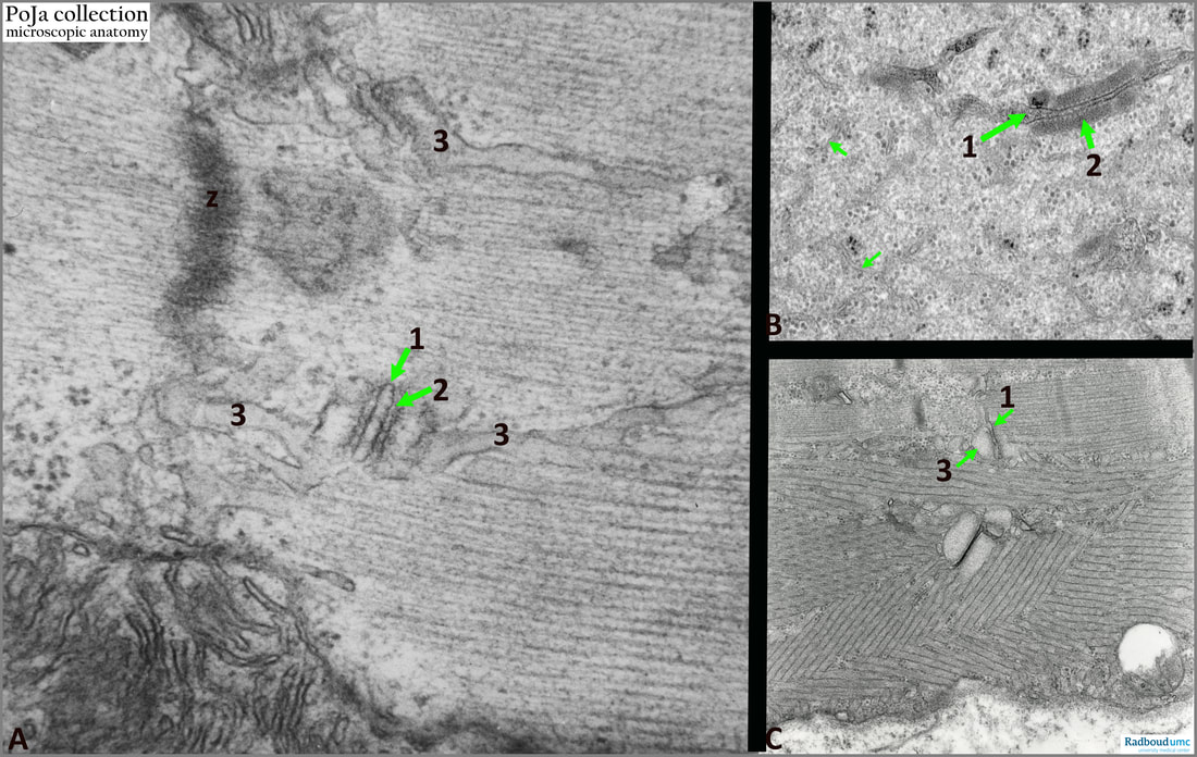
(A): Normal mouse triad with (1) T-tubule and on both sites the terminal cisternae of the sarcoplasmic reticulum (3). (2): points to the spikes or “feet” (complex of ryanodine receptors (RyR1) being the ‘connection’ between two terminal cisterns and the T-tubule. Note the dense Z-line or Z- disc.
(B): The two terminal cisterns are stained densely (2) and associate with the T-tubule (1). “Feet” are well visible. The image shows a network of thin sarcoplasmic reticulum cisternae (small arrows) and dispersed glycogen granules. Here a disturbance of the contractile apparatus resulted in a lack of myofilaments (human).
(C): The image shows the human triad with pathological hyperplasia of the sarcoplasmic reticulum, i.e., aggregates of numerous parallel arranged SER-derived tubules. (1): T-tubule. (3): Terminal cisternae. These tubular aggregates are frequently found in disorders characterised by periodic paralysis associated with disturbances in serum potassium level, both hypo- and hyperkalaemia.
See also:
Keywords/Mesh: locomotor system, skeletal muscle, striated muscle, myofibre, Z-line, sarcoplasmic reticulum (SER), T-tubule, triad, ryanodine receptor, hyperplasia SER, electron microscopy, pathology, POJA collection
Title: Ultrastructure of triad system in skeletal muscle fibres
Description:
(A): Normal mouse triad with (1) T-tubule and on both sites the terminal cisternae of the sarcoplasmic reticulum (3). (2): points to the spikes or “feet” (complex of ryanodine receptors (RyR1) being the ‘connection’ between two terminal cisterns and the T-tubule. Note the dense Z-line or Z- disc.
(B): The two terminal cisterns are stained densely (2) and associate with the T-tubule (1). “Feet” are well visible. The image shows a network of thin sarcoplasmic reticulum cisternae (small arrows) and dispersed glycogen granules. Here a disturbance of the contractile apparatus resulted in a lack of myofilaments (human).
(C): The image shows the human triad with pathological hyperplasia of the sarcoplasmic reticulum, i.e., aggregates of numerous parallel arranged SER-derived tubules. (1): T-tubule. (3): Terminal cisternae. These tubular aggregates are frequently found in disorders characterised by periodic paralysis associated with disturbances in serum potassium level, both hypo- and hyperkalaemia.
See also:
- 14.1 POJA-L6296-B Scheme excitation-contraction coupling at the Triad junction
- 14.1 POJA-L6057+6059+6060+6061 T-tubules and triads in striated muscle
- 14.1 POJA-L6248 Electron micrograph of the triad structure in skeletal muscle
- 14.1 POJA-L6229 Electron micrograph of striated muscle: triads
Keywords/Mesh: locomotor system, skeletal muscle, striated muscle, myofibre, Z-line, sarcoplasmic reticulum (SER), T-tubule, triad, ryanodine receptor, hyperplasia SER, electron microscopy, pathology, POJA collection

