
2.2 POJA –L1011. Phagocytosis in small splenic blood vessel (mouse)
2.2 POJA –L1011
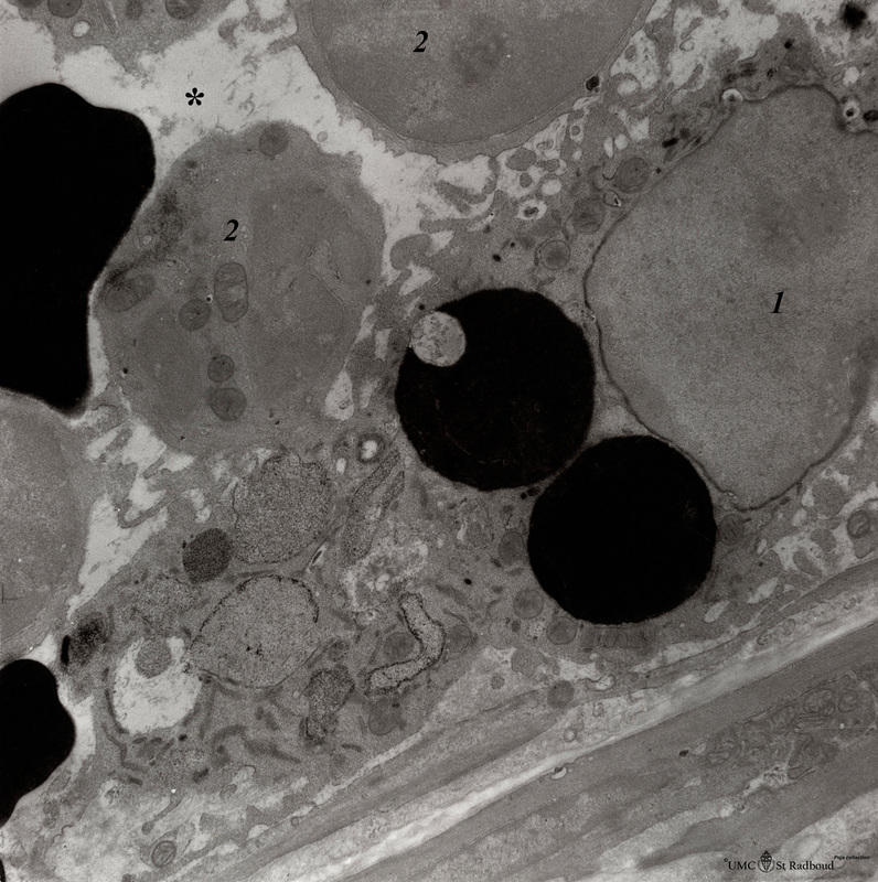
Title: Phagocytosis in small splenic blood vessel (mouse)
Description: Electron microscopy. Stain: peroxidase reaction with diaminobenzidin staining.
A diversity of red blood cells is black-stained due to the staining of haemoglobin by oxidized benzidin. Circulating lymphocytes (2) in the lumen (*) remain unstained. An oblong monocyte (1) developing into a macrophage has already ingested two black-stained erythrocytes. The cell also contains stippled large lysosomal vacuoles.
Note that the developing macrophage (1) contains dark-stained small granules but still shows negative reacting profiles of endoplasmic reticulum. The free side of the phagocyte shows finger like podia into the lumen. The underlying endothelial lining as well as few smooth muscle cells are negative.
Keywords/Mesh: lymphatic tissue, spleen, phagocytosis, histology, electron microscopy, POJA collection