
11.5 POJA-L3019+2971+
3084+3051.
Nissl bodies in neurons of cerebrum
11.5 POJA-L3019+2971+3084+3051
Title: Nissl bodies in neurons of cerebrum
Description:
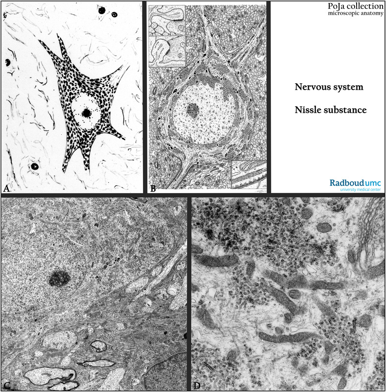
(A): Scheme of a multipolar nerve cell, human. The Nissl bodies in the cytoplasm are stained black as aggregates of mainly RER and free ribosomes.
(B): Electron microscopy scheme of large neuron with abundant aggregates of Nissl bodies, e.g. cerebellar Purkinje cell, human.
(C, D): Electron micrographs of a neuron in the substantia nigra here embedded in neuropil of glia cells and nerve fibers, rat.
The myelinated ones are recognizable as densely stained wraps around the clear axons. The neuron is rich in Nissl bodies, detailed in (D) and contained electron-dense pigment granules.
Background: In the midbrain the substantia nigra as an important motor centre is found dorsal close to the cerebral peduncles. It is involved as a lesion site in paralysis agitans (Parkinson’s disease).
Keywords/Mesh: nervous tissue, multipolar nerve cell, neuron, glia cell, neuropil, axon, Nissl body, histology, electron microscopy, POJA collection
Title: Nissl bodies in neurons of cerebrum
Description:
(A): Scheme of a multipolar nerve cell, human. The Nissl bodies in the cytoplasm are stained black as aggregates of mainly RER and free ribosomes.
(B): Electron microscopy scheme of large neuron with abundant aggregates of Nissl bodies, e.g. cerebellar Purkinje cell, human.
(C, D): Electron micrographs of a neuron in the substantia nigra here embedded in neuropil of glia cells and nerve fibers, rat.
The myelinated ones are recognizable as densely stained wraps around the clear axons. The neuron is rich in Nissl bodies, detailed in (D) and contained electron-dense pigment granules.
Background: In the midbrain the substantia nigra as an important motor centre is found dorsal close to the cerebral peduncles. It is involved as a lesion site in paralysis agitans (Parkinson’s disease).
Keywords/Mesh: nervous tissue, multipolar nerve cell, neuron, glia cell, neuropil, axon, Nissl body, histology, electron microscopy, POJA collection